Randomized Controlled Trial
. 2023:37:103313.
doi: 10.1016/j.nicl.2023.103313. Epub 2023 Jan 3.

Increased top-down control of emotions during symptom provocation working memory tasks following a RCT of alpha-down neurofeedback in PTSD

Affiliations
Randomized Controlled Trial

Increased top-down control of emotions during symptom provocation working memory tasks following a RCT of alpha-down neurofeedback in PTSD

Saurabh Bhaskar Shaw et al. Neuroimage Clin. 2023.

Abstract

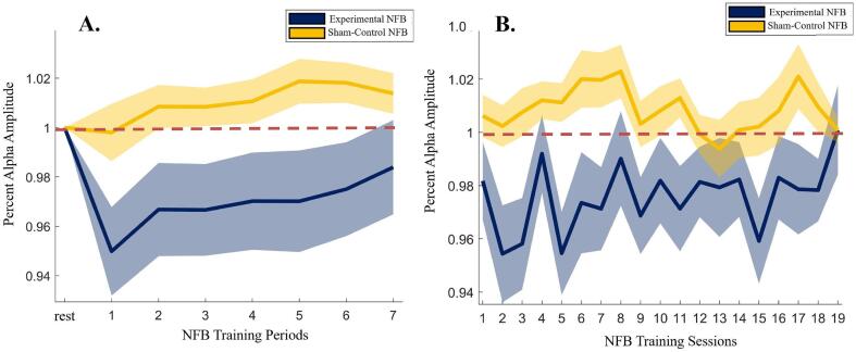
Background: Posttraumatic stress disorder (PTSD) has been found to be associated with emotion under-modulation from the prefrontal cortex and a breakdown of the top-down control of cognition and emotion. Novel adjunct therapies such as neurofeedback (NFB) have been shown to normalize aberrant neural circuits that underlie PTSD psychopathology at rest. However, little evidence exists for NFB-linked neural improvements under emotionally relevant cognitive load. The current study sought to address this gap by examining the effects of alpha-down NFB in the context of an emotional n-back task.

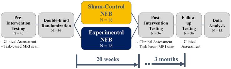
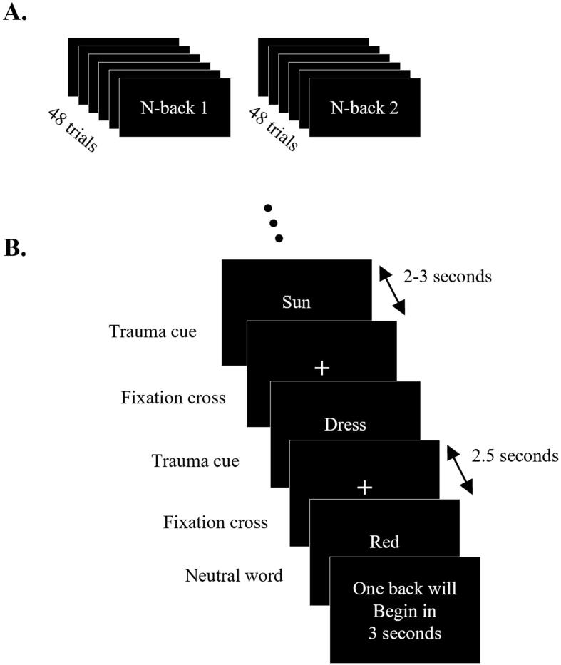
Methods: We conducted a 20-week double-blind randomized, sham-controlled trial of alpha-down NFB and collected neuroimaging data before and after the NFB protocol. Participants performed an emotional 1-back and 2-back working memory task, with interleaved trauma-neutral and trauma-relevant cues in the fMRI scanner. Data from 35 participants with a primary diagnosis of PTSD were analyzed in this study (n = 18 in the experimental group undergoing alpha-down NFB, n = 17 in the sham-control group).

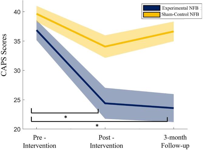
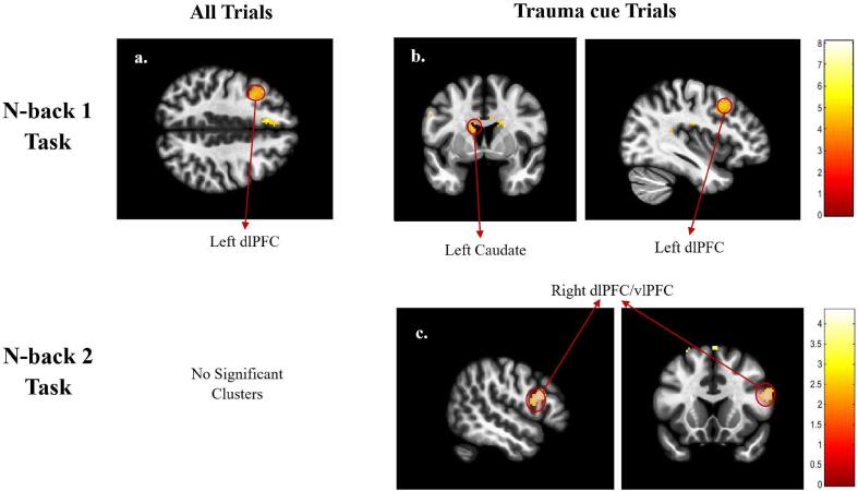
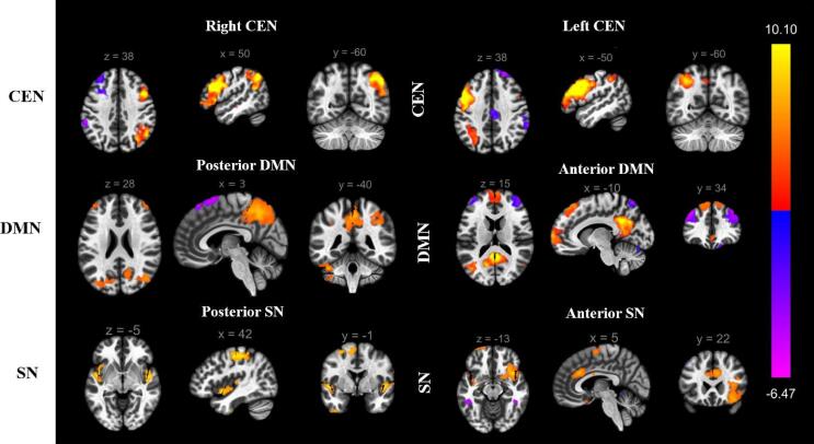
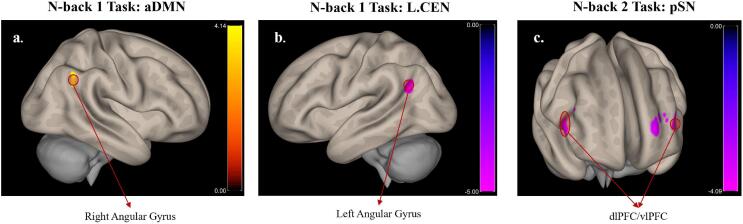
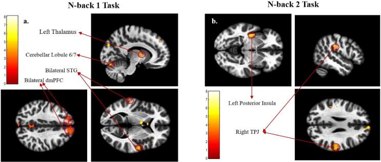
Results: Firstly, within-group analyses showed clinically significant reductions in PTSD symptom severity scores at the post-intervention timepoint and 3-month follow-up for the experimental group, and not for the sham-control group. The neuroimaging analyses revealed that alpha-down NFB enhanced engagement of top-down cognitive and emotional control centers, such as the dorsolateral prefrontal cortex (dlPFC), and improved integration of the anterior and posterior parts of the default mode network (DMN). Finally, our results also indicate that increased alpha-down NFB performance correlated with increased activity in brain regions involved in top-down control and bodily consciousness/embodied processing of self (TPJ and posterior insula).

Conclusion: This is the first study to provide mechanistic insights into how NFB may normalize dysfunctional brain activity and connectivity in PTSD under cognitive load with simultaneous symptom provocation, adding to a growing body of evidence supporting the therapeutic neuromodulatory effects of NFB. This preliminary study highlights the benefits of alpha-down NFB training as an adjunctive therapy for PTSD and warrants further investigation into its therapeutic effects on cognitive and emotion control in those with PTSD.

Keywords: Cognition; DLPFC; Emotion regulation; Functional magnetic resonance imaging (fMRI); Neurofeedback (NFB); Post-traumatic stress disorder (PTSD); Top-down control.


Conflict of interest statement

Declaration of Competing Interest The authors declare that they have no known competing financial interests or personal relationships that could have appeared to influence the work reported in this paper.

Figures

Fig. 1
Fig. 1
Schematic of the study design showing the number of participants through each section of the study.
Fig. 2
Fig. 2
Schematic of the n-back 1 and n-back 2 tasks. A. shows the two tasks, while B. shows the order and timings of the trials within each task. The trauma and neutral word cues were presented in a random order. For the n-back 1 task, the participants were instructed to press a button if the current word was identical to the previous word, while for the n-back 2 task the participants were instructed to press a button if the current word was identical to the word shown two words ago.
Fig. 3
Fig. 3
CAPS score for the experimental NFB (blue) and Sham (yellow) groups at the pre-intervention, post-intervention, and the 3-month follow-up timepoints. The shaded regions represent ± 1 SE. (For interpretation of the references to color in this figure legend, the reader is referred to the web version of this article.)
Fig. 4
Fig. 4
Average alpha amplitude at the site of NFB training (Pz electrode), shown for each of the A. seven NFB training periods within each 20-minute session (averaged across all sessions), and B. NFB training sessions (averaged across all periods). Note that the alpha amplitudes are expressed as percentage change with respect to the 3-minute “rest” period at the start of each session. Therefore, a value < 1 represents a decrease in alpha amplitude relative to the “rest” period, a value > 1 represents an increase in alpha amplitude relative to the “rest” period, and a value of 1 represents no change in the alpha amplitude from the “rest” period (represented by the dashed red line). The shaded regions represent ± 1 SE. (For interpretation of the references to color in this figure legend, the reader is referred to the web version of this article.)
Fig. 5
Fig. 5
Whole-brain activation results of the contrast experimental NFB > sham & post-intervention > pre-intervention, shown for all trials in the first column, and the Trauma cue trials in the following columns). These are shown for the n-back 1 task (first row) and the n-back 2 task (second row). The Neutral cue trials are not shown since no significant clusters were found within these trials. Significant clusters of activation are labeled. The colors represent the T-values of the voxels for the given contrast, as shown in the color bars. Abbreviations: dlPFC - Dorsolateral Prefrontal Cortex, vlPFC - Ventrolateral Prefrontal Cortex.
Fig. 6
Fig. 6
Intrinsic Connectivity Networks (ICNs) identified using independent component analysis (ICA). The labels are assigned based on spatial similarity to the networks identified in Shirer et al., 2012. For the sake of consistency with recent literature, the ventral DMN and dorsal DMN defined in Shirer et al. (2012) have been renamed posterior DMN and anterior DMN, respectively. Abbreviations: CEN - Central Executive Network, DMN - Default Mode Network, SN - Salience Network.
Fig. 7
Fig. 7
Connectivity of ICA networks with other brain regions, shown for the contrast experimental NFB > Sham & Post-intervention > Pre-intervention. The images were created by first thresholding the data at p(uncorr) < 0.001, and then identifying the significant clusters, i.e. p(FDR) < 0.05. Only the significant clusters are shown in this figure. The colors represent the T-values of the voxels for the given contrast, as shown in the color bars. Abbreviations: CEN - Central Executive Network, aDMN - Anterior Default Mode Network, pSN - Posterior Salience Network, dlPFC - Dorsolateral Prefrontal Cortex, vlPFC - Ventrolateral Prefrontal Cortex.
Fig. 8
Fig. 8
Results of the regression analysis with alpha power metric within the experimental NFB group (no significant clusters were found within the Sham group). Significant clusters that negatively correlated with alpha power metric are labeled. NOTE: due to the way the alpha power metric is defined (change in alpha power per session averaged over all NFB sessions) for the alpha-down NFB protocol, a successful NFB performer with an overall lower average alpha amplitude would correspond to a lower alpha power metric (decrease in average alpha power). Hence these clusters positively correlate with the participants’ NFB performance. The colors represent the T-values of the voxels for the given contrast, as shown in the color bars. Abbreviations: STG - Superior Temporal Gyrus, dmPFC - Dorsomedial Prefrontal Cortex, TPJ - Temporo-Parietal Junction.

References

    1. Akiki T.J., Averill C.L., Abdallah C.G. A Network-Based Neurobiological Model of PTSD: Evidence From Structural and Functional Neuroimaging Studies. Curr Psychiatry Rep. 2017;19 doi: 10.1007/s11920-017-0840-4. - DOI - PMC - PubMed
    1. Akiki T.J., Averill C.L., Wrocklage K.M., Scott J.C., Averill L.A., Schweinsburg B., Alexander-Bloch A., Martini B., Southwick S.M., Krystal J.H., Abdallah C.G. Default mode network abnormalities in posttraumatic stress disorder: A novel network-restricted topology approach. Neuroimage. 2018;176:489–498. doi: 10.1016/j.neuroimage.2018.05.005. - DOI - PMC - PubMed
    1. Ashburner J., Friston K.J. Unified segmentation. Neuroimage. 2005;26:839–851. doi: 10.1016/J.NEUROIMAGE.2005.02.018. - DOI - PubMed
    1. Aupperle R.L., Allard C.B., Grimes E.M., Simmons A.N., Flagan T., Behrooznia M., Cissell S.H., Twamley E.W., Thorp S.R., Norman S.B., Paulus M.P., Stein M.B. Dorsolateral prefrontal cortex activation during emotional anticipation and neuropsychological performance in posttraumatic stress disorder. Arch Gen Psychiatry. 2012;69:360–371. doi: 10.1001/archgenpsychiatry.2011.1539. - DOI - PubMed
    1. Barredo J., Aiken E., van 't Wout-Frank M., Greenberg B.D., Carpenter L.L., Philip N.S. Network functional architecture and aberrant functional connectivity in post-traumatic stress disorder: A clinical application of network convergence. Brain Connect. 2018;8(9):549–557. - PubMed

Publication types