. 2023 Jan 20;13(1):1124.
doi: 10.1038/s41598-023-28068-3.

Human osteoarthritis knee joint synovial fluids cleave and activate Proteinase-Activated Receptor (PAR) mediated signaling

Affiliations

Human osteoarthritis knee joint synovial fluids cleave and activate Proteinase-Activated Receptor (PAR) mediated signaling

Arundhasa Chandrabalan et al. Sci Rep. .

Abstract

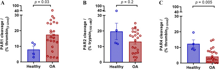
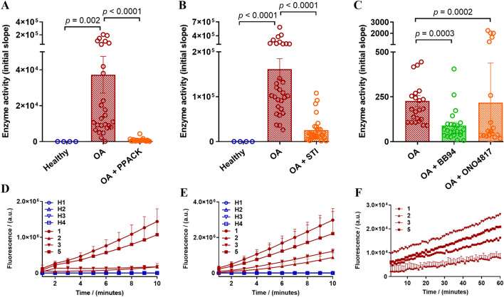
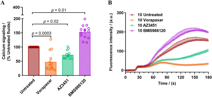
Osteoarthritis (OA) is the most prevalent joint disorder with increasing worldwide incidence. Mechanistic insights into OA pathophysiology are evolving and there are currently no disease-modifying OA drugs. An increase in protease activity is linked to progressive degradation of the cartilage in OA. Proteases also trigger inflammation through a family of G protein-coupled receptors (GPCRs) called the Proteinase-Activated Receptors (PARs). PAR signaling can trigger pro-inflammatory responses and targeting PARs is proposed as a therapeutic approach in OA. Several enzymes can cleave the PAR N-terminus, but the endogenous protease activators of PARs in OA remain unclear. Here we characterized PAR activating enzymes in knee joint synovial fluids from OA patients and healthy donors using genetically encoded PAR biosensor expressing cells. Calcium signaling assays were performed to examine receptor activation. The class and type of enzymes cleaving the PARs was further characterized using protease inhibitors and fluorogenic substrates. We find that PAR1, PAR2 and PAR4 activating enzymes are present in knee joint synovial fluids from healthy controls and OA patients. Compared to healthy controls, PAR1 activating enzymes are elevated in OA synovial fluids while PAR4 activating enzyme levels are decreased. Using enzyme class and type selective inhibitors and fluorogenic substrates we find that multiple PAR activating enzymes are present in OA joint fluids and identify serine proteinases (thrombin and trypsin-like) and matrix metalloproteinases as the major classes of PAR activating enzymes in the OA synovial fluids. Synovial fluid driven increase in calcium signaling was significantly reduced in cells treated with PAR1 and PAR2 antagonists, but not in PAR4 antagonist treated cells. OA associated elevation of PAR1 cleavage suggests that targeting this receptor may be beneficial in the treatment of OA.


Conflict of interest statement

The authors declare no competing interests.

Figures

Figure 1
Figure 1
Characterization of PAR biosensor expressing reporter cell lines, (A) nLuc-hPAR1-eYFP-CHO response to thrombin (0.003–3 U ml−1), (B) nLuc-hPAR2-eYFP-CHO response to trypsin (0.1–100 nM), and (C) nLuc-hPAR4-eYFP-CHO response to (i) thrombin (0.01–10 U ml−1) and (ii) trypsin (0.1–100 nM). Each data point on the concentration-effect curve represents the mean ± SEM of at least three independent experiments (N = 3–4) performed in triplicate.
Figure 2
Figure 2
Cleavage of PARs by enzymes in human OA knee joint synovial fluids. Healthy and OA patient synovial fluids (10%) were applied to nLuc-hPAR-eYFP-CHO cells and release of the nLuc tag into the culture supernatants was monitored as an index of receptor N-terminus cleavage. (A) Cleavage of PAR1 by synovial fluids as a % of 3 U ml−1 thrombin cleavage, (B) Cleavage of PAR2 by synovial fluids as a % of 100 nM trypsin cleavage, and (C) Cleavage of PAR4 as a % of 3 U ml−1 thrombin cleavage. Histogram represents the mean cleavage ± SEM (N = 3–5) performed in triplicate. Mann–Whitney U test was utilized to assess differences between groups. p < 0.05 was considered statistically significant.
Figure 3
Figure 3
Effect of protease inhibitors on PAR cleavage by eight human OA knee joint synovial fluids. Standard PAR activating enzymes (thrombin for PAR1 and trypsin for PAR2) or synovial fluids (10%) were pretreated with an enzyme-selective inhibitor, PPACK, STI or BB-94, before addition to nLuc-hPAR-eYFP-CHO cells. Release of the N-terminal nLuc tag into the culture supernatants was monitored as an index of receptor N-terminus cleavage. (A) PAR1 cleavage and (B) PAR2 cleavage. Each bar represents the mean ± SEM (N = 3–6). One-Way ANOVA Kruskal–Wallis test was utilized to assess differences between groups. p < 0.05 was considered statistically significant.
Figure 4
Figure 4
Cleavage of enzyme class preferred fluorogenic substrates by healthy and OA knee joint synovial fluids. Fluorogenic substrates were incubated with the healthy and OA synovial fluids (10%) with and without enzyme inhibitors (PPACK, STI, BB-94 or ONO-4817) and cleavage of (A) Bz-FVR-AMC (thrombin-like enzymes), (B) Boc-QAR-AMC (trypsin-like enzymes), and (C) MCA-KPLGL-Dpa(DNP)-AR-NH2 (MMPs) substrates was monitored. Representative kinetic traces of four healthy and four patient samples obtained with (D) Bz-FVR-AMC (10 min) and (E) Boc-QAR-AMC (10 min), and (F) four patient samples with MCA-KPLGL-Dpa(DNP)-AR-NH2 (60 min) substrates. The data represents the mean ± SEM (N = 1–3). One-Way ANOVA Kruskal–Wallis test was utilized to assess differences between groups. p < 0.05 was considered statistically significant.
Figure 5
Figure 5
OA joint fluid activation of PAR mediated calcium signaling. Synovial fluids (10%) were added to HEK293 cells expressing PAR1, PAR2 and PAR4. Elevation in intracellular calcium levels were monitored in (A) untreated cells and cells pretreated with PAR antagonists, Vorapaxar, 1000 nM (PAR1 antagonist), AZ3451, 1000 nM (PAR2 antagonist) and BMS986120, 1000 nM (PAR4 antagonist). Scatter plots depict peak calcium signaling levels in individual experiments as a percentage of the response elicited by the synovial fluid samples in untreated cells. One-Way ANOVA Kruskal–Wallis test was utilized to assess differences between groups. p < 0.05 was considered statistically significant. (B) A representative calcium signaling trace obtained for PAR mediated calcium signaling triggered by one patient sample in the absence and presence of PAR1, PAR2, and PAR4 antagonists. The data represent the mean ± SEM of three independent experiments (N = 3) performed in duplicate.

References

    1. Safiri S, et al. Global, regional and national burden of osteoarthritis 1990–2017: A systematic analysis of the Global Burden of Disease Study 2017. Ann. Rheum. Dis. 2020;79:819–828. doi: 10.1136/annrheumdis-2019-216515. - DOI - PubMed
    1. Hunter DJ, Bierma-Zeinstra S. Osteoarthritis. Lancet Lond. Engl. 2019;393:1745–1759. doi: 10.1016/S0140-6736(19)30417-9. - DOI - PubMed
    1. Kloppenburg M, Berenbaum F. Osteoarthritis year in review 2019: Epidemiology and therapy. Osteoarthritis Cartilage. 2020;28:242–248. doi: 10.1016/j.joca.2020.01.002. - DOI - PubMed
    1. Rengel Y, Ospelt C, Gay S. Proteinases in the joint: Clinical relevance of proteinases in joint destruction. Arthritis Res. Ther. 2007;9:221. doi: 10.1186/ar2304. - DOI - PMC - PubMed
    1. Miller RE, Lu Y, Tortorella MD, Malfait A-M. Genetically engineered mouse models reveal the importance of proteases as osteoarthritis drug targets. Curr. Rheumatol. Rep. 2013;15:350. doi: 10.1007/s11926-013-0350-2. - DOI - PMC - PubMed

Publication types

Grants and funding