. 2023 Jan 20;13(1):1163.
doi: 10.1038/s41598-023-27959-9.

Membrane vesicles released by Lacticaseibacillus casei BL23 inhibit the biofilm formation of Salmonella Enteritidis

Affiliations

Membrane vesicles released by Lacticaseibacillus casei BL23 inhibit the biofilm formation of Salmonella Enteritidis

David da Silva Barreira et al. Sci Rep. .

Abstract

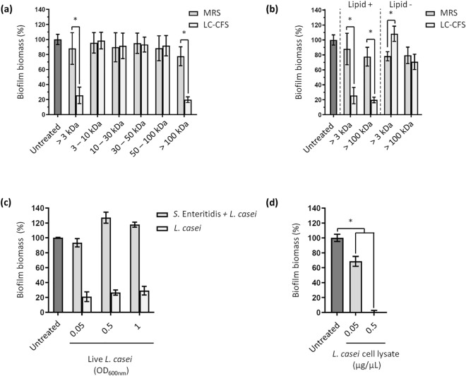
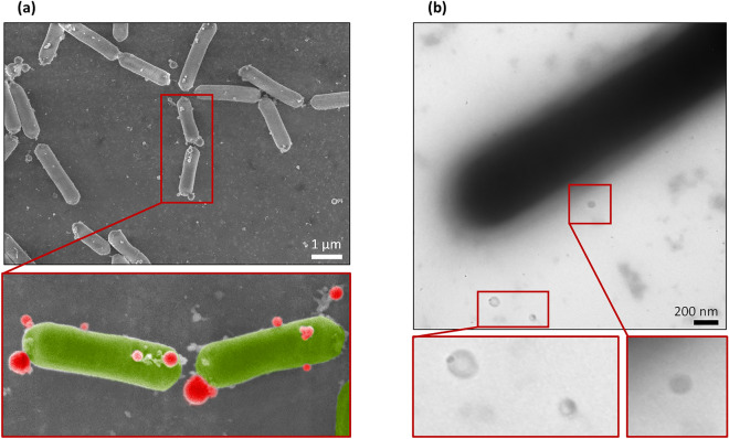
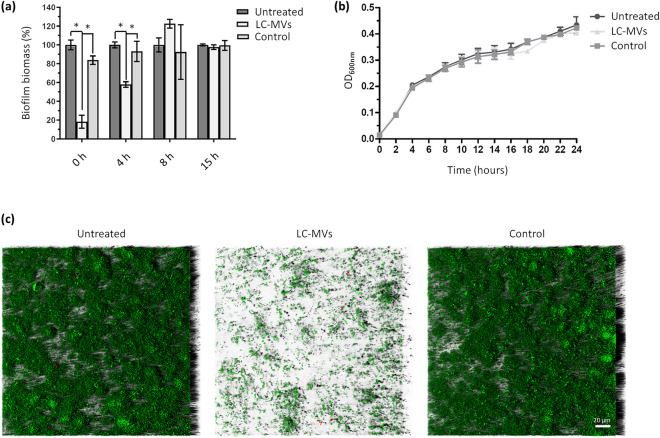
Biofilms represent a major concern in the food industry and healthcare. The use of probiotic bacteria and their derivatives as an alternative to conventional treatments to fight biofilm development is a promising option that has provided convincing results in the last decades. Recently, membrane vesicles (MVs) produced by probiotics have generated considerable interest due to the diversity of roles they have been associated with. However, the antimicrobial activity of probiotic MVs remains to be studied. In this work, we showed that membrane vesicles produced by Lacticaseibacillus casei BL23 (LC-MVs) exhibited strong antibiofilm activity against Salmonella enterica serovar Enteritidis (S. Enteritidis) without affecting bacterial growth. Furthermore, we found that LC-MVs affected the early stages of S. Enteritidis biofilm development and prevented attachment of bacteria to polystyrene surfaces. Importantly, LC-MVs did not impact the biomass of already established biofilms. We also demonstrated that the antibiofilm activity depended on the proteins associated with the LC-MV fraction. Finally, two peptidoglycan hydrolases (PGHs) were found to be associated with the antibiofilm activity of LC-MVs. Overall, this work allowed to identify the antibiofilm properties of LC-MVs and paved the way for the use of probiotic MVs against the development of negative biofilms.


Conflict of interest statement

The authors declare no competing interests.

Figures

Figure 1
Figure 1
L. casei BL23 releases lipid components of high molecular weight in the supernatant with antibiofilm properties. (a) S. Enteritidis was grown in polystyrene microplates and treated with multiple fractions of L. casei cell-free supernatant (LC-CFS) and MRS medium (MRS). After 24 h of culture, the biofilms were quantified by crystal violet staining. The LC-CFS and MRS medium were fractionated by size-exclusion ultrafiltration, generating fractions with molecular weight ranging from 3 kDa to over 100 kDa. (b) The LC-CFS fractions exhibiting an antibiofilm activity and the corresponding MRS fractions were treated with a lipid adsorption matrix to selectively remove all the lipids. Biofilm biomasses of S. Enteritidis were then quantified by crystal violet staining after treatment with the fractions in the absence (Lipid −) and in the presence (Lipid +) of lipids. (c) S. Enteritidis biofilm formation was quantified after treatment with live L. casei. Please note that the control condition “L. casei” shows the biomass formed by L. casei BL23 in TSB without S. Enteritidis. (d) S. Enteritidis biofilm formation was quantified after treatment with lysed L. casei. All the results were normalized to the untreated conditions and expressed as a percentage.
Figure 2
Figure 2
L. casei BL23 release membrane vesicles in the supernatant. SEM (a) and negative-staining TEM (b) images showing that MVs produced by L. casei BL23 are found on the surface of the bacteria and free in the supernatant. The bottom images show a magnified view of each EM images. MVs were colored in red and bacteria were colored in green in the SEM zoomed-in view for better identification.
Figure 3
Figure 3
Membrane vesicles released by L. casei BL23 (LC-MVs) inhibit the early stages of biofilm formation without impacting the growth of S. Enteritidis. (a) S. Enteritidis was grown in polystyrene microplates and treated with LC-MVs (0.04 µg/µL) or a control fraction after 0, 4, 8 and 15 h of growth. After 24 h of culture, biofilms were quantified by crystal violet staining. The control corresponds to the fraction collected after carrying out the purification protocol on the culture medium alone (i.e., MRS medium). Results were normalized to the untreated conditions and expressed as a percentage. (b) Comparison of S. Enteritidis growth curves in the absence and in the presence of treatment with LC-MVs (0.04 µg/µL) and the control fraction. (c) The biofilms formed by S. Enteritidis after 24 h of culture in the absence or in the presence of treatment with LC-MVs (0.04 µg/µl) were stained (live/dead; SYTO 9/propidium iodide) and imaged by confocal laser scanning microscopy (CLSM). Green: total biomass. Red: dead cells.
Figure 4
Figure 4
The antibiofilm activity of LC-MVs is sensitive to heat and proteinase K treatment. (a) LC-MVs and the control fraction were subjected to proteinase K treatment, heat inactivation (10 min at 70 °C and 100 °C) and lipid removal using a lipid adsorption reagent. Biofilm biomasses of S. Enteritidis were then quantified by crystal violet staining after adding the treated MVs and control fractions. (b) S. Enteritidis was incubated with increasing quantities of LC-MVs for 30 min at 37 °C before inoculation in polystyrene microplates. After 24 h of culture, the biofilm biomasses were quantified by crystal violet staining.
Figure 5
Figure 5
Mutation of two of the most abundant proteins in the vesicular fraction partially suppresses the antibiofilm activities of LC-MVs against S. Enteritidis. (a) List of the peptidoglycan hydrolases (PGHs) detected with the vesicles of L. casei BL23 and selected for mutagenesis. The column “Mutant name” indicates the names of the mutant strains obtained by the insertion of a non-replicative plasmid into genes of L. casei encoding PGHs. (b) The MVs of the mutant strains were purified and their antibiofilm activity against S. Enteritidis was analyzed, as described previously. For each condition, S. Enteritidis was treated with a final quantity of 0.04 µg/µL of MVs.
Figure 6
Figure 6
Schematic representation of the effect of LC-MVs treatment on S. Enteritidis biofilm formation. Proteins (in red) associated with LC-MVs inhibit the attachment of S. Enteritidis on polystyrene surfaces, preventing the formation of biofilm. Two peptidoglycan hydrolases (PGHs) were found to be involved in the antibiofilm activity of LC-MVs.

References

    1. Flemming H-C, Wuertz S. Bacteria and archaea on earth and their abundance in biofilms. Nat. Rev. Microbiol. 2019;17:247–260. doi: 10.1038/s41579-019-0158-9. - DOI - PubMed
    1. Flemming H-C, et al. Who put the film in biofilm? The migration of a term from wastewater engineering to medicine and beyond. NPJ Biofilms Microbiomes. 2021;7:10. doi: 10.1038/s41522-020-00183-3. - DOI - PMC - PubMed
    1. Vert M, et al. Terminology for biorelated polymers and applications (IUPAC Recommendations 2012) Pure Appl. Chem. 2012;84:377–410. doi: 10.1351/PAC-REC-10-12-04. - DOI
    1. Flemming H-C, Wingender J. The biofilm matrix. Nat. Rev. Microbiol. 2010;8:623–633. doi: 10.1038/nrmicro2415. - DOI - PubMed
    1. Elasri MO, Miller RV. Study of the response of a biofilm bacterial community to UV radiation. Appl. Environ. Microbiol. 1999;65:2025–2031. doi: 10.1128/AEM.65.5.2025-2031.1999. - DOI - PMC - PubMed

Publication types