Review
. 2023 Jan 20;9(1):3.
doi: 10.1186/s40942-023-00443-2.

Pathomechanisms in central serous chorioretinopathy: A recent update

Affiliations
Review

Pathomechanisms in central serous chorioretinopathy: A recent update

Arman Zarnegar et al. Int J Retina Vitreous. .

Abstract

Background: Central serous chorioretinopathy (CSCR) is a potentially blinding choroidal disease. Despite decades of research, the pathological mechanisms of CSCR are still poorly understood. In recent years, there has been a strong emphasis on choroidal dysfunction as a primary cause of CSCR.

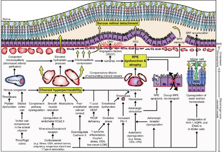
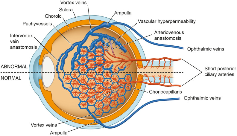
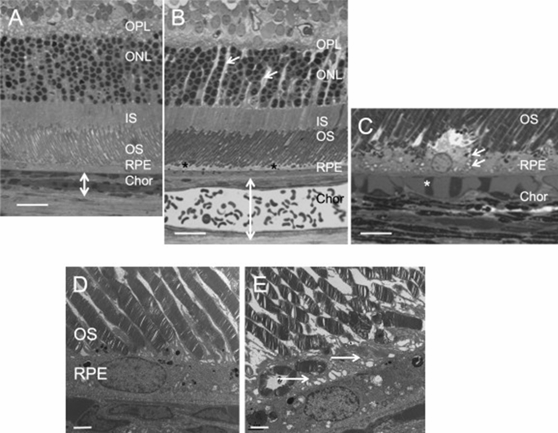
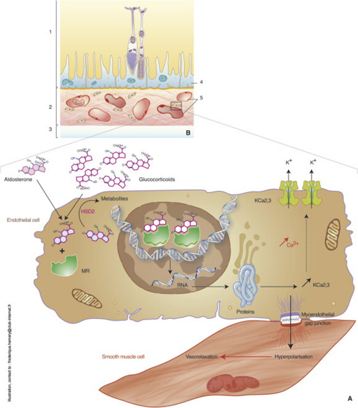
Main body: The concept of the pachychoroid disease spectrum and pachychoroid-driven processes are central to current theories regarding the pathophysiological underpinnings of CSCR. Choroidal hyperpermeability and subsequent leakage of fluid seen in CSCR may be due to several causes. Among them are venous congestion, inflammation, mineralocorticoid receptor activation, systemic factors including hemodynamic changes, obstructive sleep apnea, phosphodiesterase inhibitor use, pregnancy, and genetic predispositions. Congestion of vortex veins that drain blood from the choroid may contribute to the dilation of Haller vessels and cause fluid leakage. Vortex veins exit the eye through the sclera; thus, increased scleral thickness has been proposed to be a factor in venous congestion. Asymmetric vortex vein drainage may similarly result in congestion of the local venous system. Vortex vein anastomoses may overload the venous system and form secondary to venous congestion. Recent studies suggest inflammation and mineralocorticoid activation may factor into the development of CSCR, though more research in these areas is called for. Systemic conditions and genetics may predispose individuals to develop CSCR.

Conclusions: By striving to understand the molecular and physiological mechanisms of this disease, we can better diagnose and treat CSCR to improve outcomes for patients.

Keywords: Central serous chorioretinopathy; Choroid; Pachychoroid disease; Retina.


Conflict of interest statement

The authors declare that they have no competing interests.

Figures

Fig. 1
Fig. 1
Molecular pathophysiological mechanisms causing serous retinal detachment, pigment epithelial detachment, and choroidal neovascularization in CSCR. The upstream mechanisms leading to choroid hyperpermeability include vortex vein compression, corticosteroid receptor activation, and oxidative stress/inflammation. Reprinted with permission from Kanda et al. [66] Pathophysiology of central serous chorioretinopathy: a literature review with quality assessment. Eye 36, 941–962 (2022) under Springer Nature and Copyright Clearance Center Rights Link License
Fig. 2
Fig. 2
Indocyanine green angiography demonstrating vortex veins in the left eye of a patient with CSCR. (A) Early and (B) late phases show dilated vessels and late leakage
Fig. 3
Fig. 3
Diagram demonstrating choroidal arteriovenous vasculature. Healthy (bottom) and diseased (top) choroidal arteriovenous vasculature. In the abnormal choroidal arteriovenous vasculature, pathological anastomoses that bypass the capillary bed may form to cause an increased arterial filling in the vortex veins, leading to vortex vein congestion. This engorgement can lead to choriocapillaris congestion, leading to the release of angiogenic factors and the breakdown of the RPE as seen in CSCR. Reprinted with permission from Brinks et al. [24] Choroidal arteriovenous anastomoses: a hypothesis for the pathogenesis of central serous chorioretinopathy and other pachychoroid disease spectrum abnormalities. (2022) under Creative Commons Attribution-NonCommercial-NoDerivatives 4.0 International (CC BY-NC-ND 4.0) License (https://creativecommons.org/licenses/by-nc-nd/4.0/legalcode)
Fig. 4
Fig. 4
Swept source anterior segment OCT of the left eye of a 45-year-old male with CSCR. A color fundus photograph (A) demonstrates neurosensory retinal detachment secondary to SRF accumulation in the macula. Swept source OCT (B) shows SRF in the fovea with a subfoveal choroidal thickness of 469 μm and Haller vessel dilation. Scleral thickness was measured via anterior segment swept source OCT at nasal (C), inferior (D), temporal (E), and superior (F) regions with thicknesses of 455 μm, 485 μm, 442 μm, and 523 μm, respectively. Asterisks represent rectus muscles. Reprinted with permission from Imanaga et al. [26], under Creative Commons Attribution-NonCommercial-NoDerivatives 4.0 International (CC BY-NC-ND 4.0) License (https://creativecommons.org/licenses/by-nc-nd/4.0/legalcode)
Fig. 5
Fig. 5
Multimodal imaging of the right eye of a 62-year-old woman with pachychoroid pigment epitheliopathy. (A) Early to mid-phase ultra-widefield indocyanine green angiography exhibits asymmetric drainage within the vortex venous system. Choroidal hyperpermeability can be observed in the macula and inferonasal peripapillary areas in a late-phase ultra-widefield indocyanine green angiography (B). Choroidal thickness maps in (C) are taken from the region marked by yellow dashed boxes in (A, B). Regions of choroidal hyperpermeability from (B) also exhibit increased choroidal thickness in (C) on swept-source OCT imaging. Spectral-domain OCT of the fovea shows a nonvascularized shallowed irregular pigment epithelial detachment (D). Reprinted with permission from Bacci et al. [30], under Creative Commons Attribution-NonCommercial-NoDerivatives 4.0 International (CC BY-NC-ND 4.0) License (https://creativecommons.org/licenses/by-nc-nd/4.0/legalcode)
Fig. 6
Fig. 6
En face OCT images of the choroid. (A) Symmetrical venous drainage involving the superotemporal and inferotemporal vortex veins in a healthy eye. (B, C) Vortex vein anastomoses secondary to venous congestion in an eye with CSCR. Reprinted with permission from Spaide et al. [21], under Creative Commons Attribution-NonCommercial-NoDerivatives 4.0 International (CC BY-NC-ND 4.0) License (https://creativecommons.org/licenses/by-nc-nd/4.0/legalcode)
Fig. 7
Fig. 7
Effects of aldosterone injection in rat eyes. Control rat eyes (A, D) and aldosterone-injected rat eyes (B, C, E). Intravitreal injection of aldosterone in rat eyes in semi-thin sections shows (B) swollen Müller cells (arrows), thickened retinal pigment epithelial apical microvilli (stars), and thickened choroid (bidirectional arrows). Additionally, engorged choroidal vessels can be appreciated (C, star). Transmission electron microscopy (E) also shows an increased thickness of retinal pigment epithelial apical microvilli (arrows). INL, inner nuclear layer; IS/OS, inner and outer segments of photoreceptors. Bar: A and B, 20 μm; C, 10 μm; D and E, 2 μm. Reprinted with permission from Daruich et al. [5], under Creative Commons Attribution-NonCommercial-NoDerivatives 4.0 International (CC BY-NC-ND 4.0) License (https://creativecommons.org/licenses/by-nc-nd/4.0/legalcode)
Fig. 8
Fig. 8
Mechanisms of choroidal vascular dilation via activation of mineralocorticoid receptors. Choroidal vascular endothelial cells express mineralocorticoid receptors (MR) which can be activated by aldosterone and glucocorticoids with the downstream effect of inducing vasodilation. Reprinted with permission from Daruich et al. [5], under Creative Commons Attribution-NonCommercial-NoDerivatives 4.0 International (CC BY-NC-ND 4.0) License (https://creativecommons.org/licenses/by-nc-nd/4.0/legalcode)

References

    1. Sahoo NK, Ong J, Selvam A, Maltsev D, Sacconi R, Venkatesh R, Reddy NG, Madan S, Tombolini B, Lima LH, et al. Longitudinal follow-up and outcome analysis in central serous chorioretinopathy. Eye (Lond) 2022 doi: 10.1038/s41433-022-02044-z. - DOI - PMC - PubMed
    1. Kitzmann AS, Pulido JS, Diehl NN, Hodge DO, Burke JP. The incidence of central serous chorioretinopathy in Olmsted County, Minnesota, 1980–2002. Ophthalmology. 2008;115:169–173. doi: 10.1016/j.ophtha.2007.02.032. - DOI - PubMed
    1. Chhablani J, Cohen FB, Group CSCI. Multimodal imaging-based central serous chorioretinopathy classification. Ophthalmol Retina 2020; 4:1043–6. 10.1016/j.oret.2020.07.026. - PubMed
    1. Chhablani J, Behar-Cohen F, Group CSCI. Validation of central serous chorioretinopathy multimodal imaging-based classification system. Graefes Arch Clin Exp Ophthalmol 2022; 260:1161–9. 10.1007/s00417-021-05452-1. - PubMed
    1. Daruich A, Matet A, Dirani A, Bousquet E, Zhao M, Farman N, Jaisser F, Behar-Cohen F. Central serous chorioretinopathy: Recent findings and new physiopathology hypothesis. Prog Retin Eye Res. 2015;48:82–118. doi: 10.1016/j.preteyeres.2015.05.003. - DOI - PubMed

LinkOut - more resources