. 2023 Jan 20;11(1):11.
doi: 10.1186/s40168-022-01456-z.

The structure and diversity of microbial communities in Paederus fuscipes (Coleoptera: Staphylinidae): from ecological paradigm to pathobiome

Affiliations

The structure and diversity of microbial communities in Paederus fuscipes (Coleoptera: Staphylinidae): from ecological paradigm to pathobiome

Bahar Chamankar et al. Microbiome. .

Abstract

Background: Paederus fuscipes is medically the most famous rove beetle, which causes dermatitis or conjunctivitis in humans, as well as gastrointestinal toxicosis in livestock, via releasing toxic hemolymph containing pederin. Pedrin biosynthesis genes have been identified in uncultured Pseudomonas-like endosymbionts that are speculated to be acquired through a horizontal transfer. However, the composition of the P. fuscipes microbial community, especially of the gut and genital microbiome, remains unclear. This study was aimed to characterize the structure and diversity of P. fuscipes-associated bacterial communities in terms of gender, organ, and location using the Illumina HiSeq platform in the southern littorals of Caspian Sea.

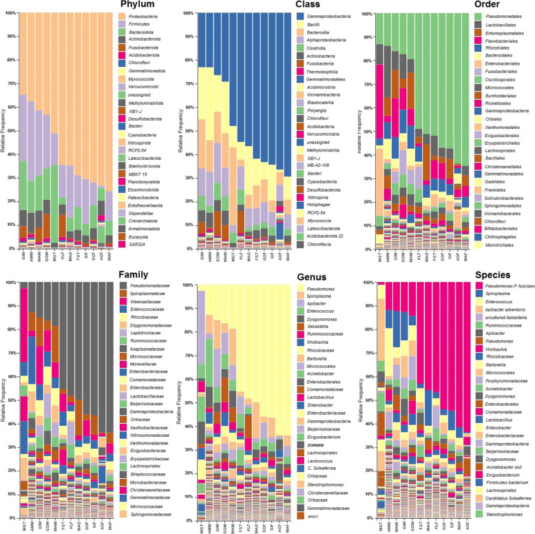
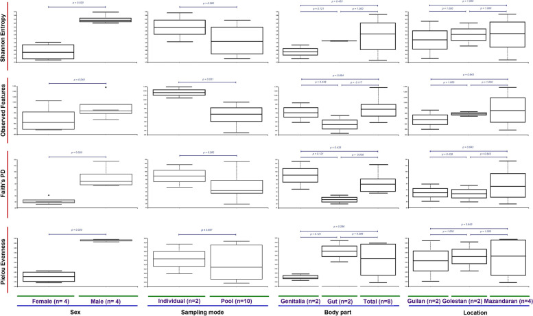
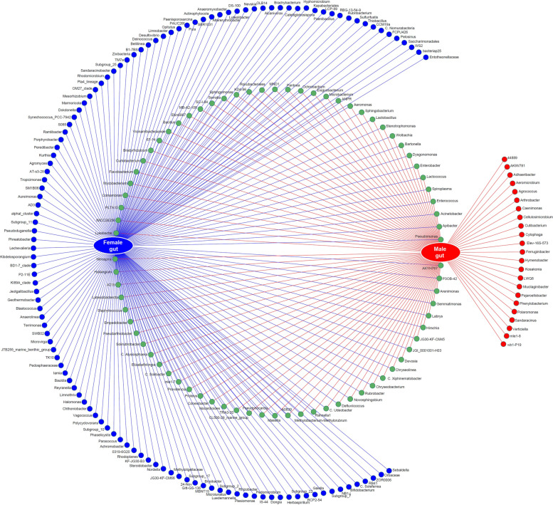
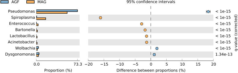
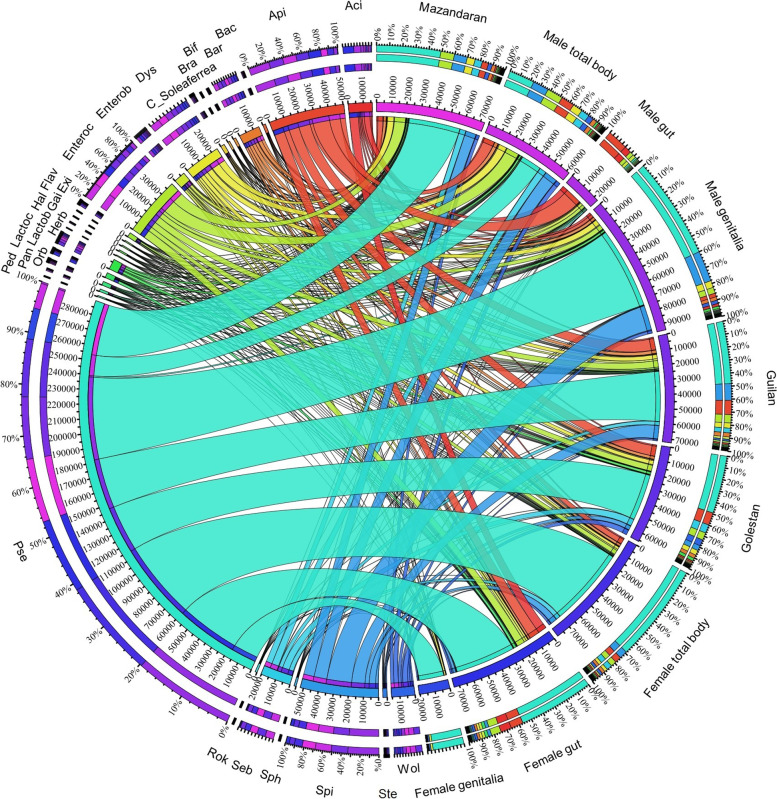
Results: The OTUs identified from P. fuscipes specimens were collapsed into 40 phyla, 112 classes, 249 orders, 365 families, 576 genera, and 106 species. The most abundant families were Pseudomonadaceae, Spiroplasmataceae, Weeksellaceae, Enterococcaceae, and Rhizobiaceae, respectively. Thirty top genera made up > 94% of the P. fuscipes microbiome, with predominating Pseudomonas, followed by the Spiroplasma, Apibacter, Enterococcus, Dysgonomonas, Sebaldella, Ruminococcus, and Wolbachia. Interesting dissimilarities were also discovered within and between the beetle microbiomes in terms of genders and organs. Analyses showed that Spiroplasma / Apibacter as well as Pseudomonas / Pseudomonas were the most abundant in the genitals / intestines of male and female beetles, respectively. Bacterial richness did not display any significant difference in the three provinces but was higher in male beetles than in females and more in the genitals than intestines.

Conclusions: The present study identified Pseudomonas-like endobacterium as a common symbiont of P. fuscipes beetles; this bacterium begins its journey from gut and genitalia of females to reach the male rove beetles. Additionally, male and female rove beetles were characterized by distinctive microbiota in different organs, likely reflecting different functions and/or adaptation processes. Evidence of the extension of P. fuscipes microbiome from the environmental paradigm to the pathobiome was also presented herein. A comprehensive survey of P. fuscipes microbiome components may eventually lead to ecological insights into the production and utilization of defensive compound of pederin and also the management of linear dermatitis with the use of available antibiotics against bacterial pathogens released by the beetles. Video Abstract.

Keywords: Apibacter; Dermatitis linearis; Genital microbiota; Gut microbiota; Pederin; Pseudomonas-like Paederus fuscipes endosymbiont; Spiroplasma; Wolbachia.


Conflict of interest statement

The authors declare that they have no competing interests.

Figures

Fig. 1
Fig. 1
Relative frequency and taxonomical composition of microbiome of Paederus fuscipes beetles by phylum, class, order, family, genus, and species. Legends are shown for 30 OTUs with high frequency
Fig. 2
Fig. 2
Box plots of alpha diversity indices (Shannon’s entropy, observed features, Faith’s PD, and Pielou’s evenness) comparing microbiome in terms of gender, sampling modes, body parts, and locality of studied Paederus fuscipes specimens
Fig. 3
Fig. 3
Principal coordinates analysis (PCoA) plots of beta diversity distances (Bray-Curtis, Jaccard, and unweighted/weighted UniFrac) between Paederus fuscipes specimens. Distances between points on the ordination plot reflect relative dissimilarities in microbiome structures. Eigenvalues of PCoA1, PCoA2, and PCoA3 are shown in parentheses. Samples were colored according to the fields sources
Fig. 4
Fig. 4
Network analysis showing the shared (green circles crossed by both red/blue lines) and non-shared gut bacteria of male (red circles) and female (blue circles) Paederus fuscipes specimens
Fig. 5
Fig. 5
Comparison of gut bacteria profile of male and female Paederus fuscipes specimens using STAMP analysis. Comparisons were made at the bacterial genus level. Corrected p values were calculated based on Fisher’s exact test method using Storey’s FDR approach. P values < 0.05 were taken for comparison. The bar plot indicated in blue or orange shows a positive or negative difference between read proportions. The size effect of >100 reads is included in the comparisons. Differences between samples are shown at 95% confidence intervals
Fig. 6
Fig. 6
Network analysis showing the shared (green circles crossed by both red/blue lines) and non-shared bacteria of male (red circles) and female (blue circles) genital tract of Paederus fuscipes
Fig. 7
Fig. 7
Comparison of genital bacteria profile of male and female Paederus fuscipes specimens using STAMP analysis. Comparisons were made at the bacterial genus level. Corrected p values were calculated based on Fisher’s exact test method using Storey’s FDR approach. P values < 0.05 were taken for comparison. The bar plot indicated in blue or orange shows a positive or negative difference between read proportions. The size effect of >100 reads is included in the comparisons. Differences between samples are shown at 95% confidence intervals
Fig. 8
Fig. 8
Principal component analysis (PCA) of the abundance of bacterial communities in the total bodies of male and females Paederus fuscipes. Bacteria from male (orange squares) beetles are fully grouping separated than female (blue circles) beetles
Fig. 9
Fig. 9
Box plot analysis of 12 bacterial families of Pseudomonadaceae, Spiroplasmataceae, Weeksellaceae, Enterococcaceae, Rhizobiaceae, Dysgonomonadaceae, Leptotrichiaceae, Ruminococcaceae, Anaplasmataceae, Micrococcaceae, Moraxellaceae, and Enterococcaceae with high abundance in the total bodies of female and male Paederus fuscipes
Fig. 10
Fig. 10
Network analysis representing the shared and exclusive bacterial genera identified from body parts and genders of Paederus fuscipes beetles captured from three Southern Caspian Sea Provinces, Guilan, Mazandaran, and Golestan. Only the top 27 bacteria in relative frequencies are shown. Aci, Acinetobacter; Api, Apibacter; Bac, Bacillus; Bar, Bartonella; Bif, Bifidobacterium; Bra, Bradyrhizobium; C_Soleaferrea, Candidatus_Soleaferrea; Dys, Dysgonomonas; Enterob, Enterobacter; Enteroc, Enterococcus; Exi, Exiguobacterium; Flav, Flavobacterium; Gai, Gaiella; Hal, Haliangium; Herb, Herbaspirillum; Lactob, Lactobacillus; Lactoc, Lactococcus; Orb, Orbaceae; Pan, Pantoea; Ped, Pedomicrobium; Pse, Pseudomonas; Rok, Rokubacteriales; Seb, Sebaldella; Sph, Sphingomonas; Spi, Spiroplasma; Ste, Stenotrophomonas; Wol, Wolbachia
Fig. 11
Fig. 11
Venn diagram representing the distribution of the bacteria genera across the body parts of the specimens from Mazandaran (left) and Mazandaran-Guilan-Golestan Provinces (right) of the Paederus fuscipes. Shared bacteria genera are shown in core. The percentage of bacteria genera for each partner is indicated within parentheses

References

    1. Basset Y, Cizek L, Cuénoud P, Didham RK, Guilhaumon F, Missa O, Novotny V, Ødegaard F, Roslin T, Schmidl J, et al. Arthropod diversity in a tropical forest. Science. 2012;338:1481–1484. doi: 10.1126/science.1226727. - DOI - PubMed
    1. van Huis A. The global impact of insects. Wageningen University, Wageningen UR; 2014.
    1. Khanzadeh F, Khaghaninia S, Maleki-Ravasan N, Oshaghi MA, Adler PH. Black flies (Diptera: Simuliidae) of the Aras River Basin: species composition and floral visitation. Acta Trop. 2020;209:105536. doi: 10.1016/j.actatropica.2020.105536. - DOI - PubMed
    1. Namaki-Khameneh R, Khaghaninia S, Disney RHL, Maleki-Ravasan N. The scuttle flies (Diptera: Phoridae) of Iran with the description of Mahabadphora aesthesphora as a new genus and species. PLoS One. 2021;16:e0257899. doi: 10.1371/journal.pone.0257899. - DOI - PMC - PubMed
    1. Maleki-Ravasan N, Oshaghi MA, Afshar D, Arandian MH, Hajikhani S, Akhavan AA, Yakhchali B, Shirazi MH, Rassi Y, Jafari R, et al. Aerobic bacterial flora of biotic and abiotic compartments of a hyperendemic Zoonotic Cutaneous Leishmaniasis (ZCL) focus. Parasites Vectors. 2015;8:1–22. doi: 10.1186/s13071-014-0517-3. - DOI - PMC - PubMed

Publication types

LinkOut - more resources