

A 5-Year-Old Child with a Deep Neck Abscess Complicated by Laryngeal Obstruction
- PMID: 36670568
- PMCID: PMC9856470
- DOI: 10.3390/children10010017
A 5-Year-Old Child with a Deep Neck Abscess Complicated by Laryngeal Obstruction
Abstract
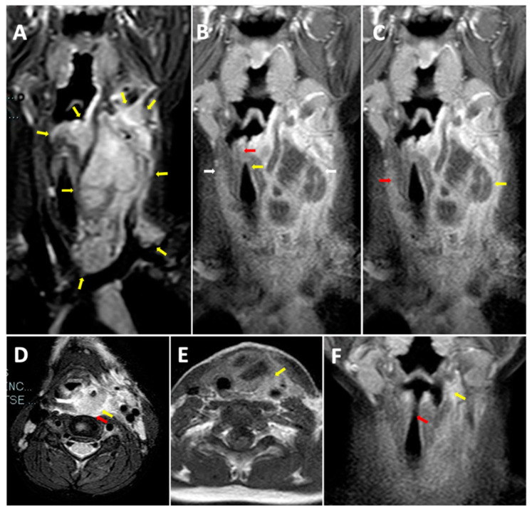
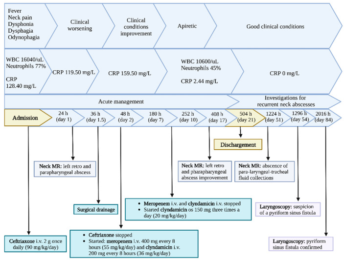
Deep neck space infections (DNSI) are defined as infections in the potential spaces and fascial planes of the neck. We show the clinical case of a retro and para-pharyngeal abscess in a healthy 5-year-old child complicated by compression and dislocation of the larynx with marked airway caliber reduction and potentially fatal extension up to the mediastinal aditus. DNSI can occur at any age and, due to its rapid progression, requires immediate treatment in children. In healthy children, concurrent abscesses in separate neck spaces are rare. DNSI recurrence should alert the physician to the possibility of a congenital problem, and if imaging fails, laryngoscopy may be the best diagnostic technique.
Keywords: deep neck space infection; neck abscess; neck pain; torticollis.
Conflict of interest statement
The authors declare no conflict of interest.
Figures
Similar articles
-
Deep Neck Space Infections: A Study of 76 Cases.Iran J Otorhinolaryngol. 2015 Jul;27(81):293-9. Iran J Otorhinolaryngol. 2015. PMID: 26788478 Free PMC article.
-
Controversies in the management of deep neck space infection in children: an evidence-based review.Clin Otolaryngol. 2017 Feb;42(1):156-163. doi: 10.1111/coa.12692. Epub 2016 Jun 30. Clin Otolaryngol. 2017. PMID: 27288654 Review.
-
Odontogenic versus nonodontogenic deep neck space infections: CT manifestations.J Comput Assist Tomogr. 1997 Mar-Apr;21(2):202-8. doi: 10.1097/00004728-199703000-00006. J Comput Assist Tomogr. 1997. PMID: 9071285
-
Bilateral deep neck space infection in pediatric patients: review of literature and report of a case.J Indian Soc Pedod Prev Dent. 2015 Jan-Mar;33(1):61-5. doi: 10.4103/0970-4388.149009. J Indian Soc Pedod Prev Dent. 2015. PMID: 25572377 Review.
-
A Study on Deep Neck Space Infections.Indian J Otolaryngol Head Neck Surg. 2019 Oct;71(Suppl 1):912-917. doi: 10.1007/s12070-019-01583-4. Epub 2019 Jan 21. Indian J Otolaryngol Head Neck Surg. 2019. PMID: 31742093 Free PMC article.
References
-
- Yellon R.F., Bluestone C.D., Casselbrant M.L., Stool S.E., GonzÁAlez-Valdepena H., Bluestone C.D. Paediatric Otolaryngology. 24th ed. Saunders; Philadelphia, PA, USA: 2003. Head and neck space infections; pp. 1681–1701.
Publication types
LinkOut - more resources
Full Text Sources
