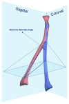
Accuracy of 3D Corrective Osteotomy for Pediatric Malunited Both-Bone Forearm Fractures
- PMID: 36670572
- PMCID: PMC9856311
- DOI: 10.3390/children10010021
Accuracy of 3D Corrective Osteotomy for Pediatric Malunited Both-Bone Forearm Fractures
Abstract
Re-displacement of a pediatric diaphyseal forearm fracture can lead to a malunion with symptomatic impairment in forearm rotation, which may require a corrective osteotomy. Corrective osteotomy with two-dimensional (2D) radiographic planning for malunited pediatric forearm fractures can be a complex procedure due to multiplanar deformities. Three-dimensional (3D) corrective osteotomy can aid the surgeon in planning and obtaining a more accurate correction and better forearm rotation. This prospective study aimed to assess the accuracy of correction after 3D corrective osteotomy for pediatric forearm malunion and if anatomic correction influences the functional outcome. Our primary outcome measures were the residual maximum deformity angle (MDA) and malrotation after 3D corrective osteotomy. Post-operative MDA > 5° or residual malrotation > 15° were defined as non-anatomic corrections. Our secondary outcome measure was the gain in pro-supination. Between 2016−2018, fifteen patients underwent 3D corrective osteotomies for pediatric malunited diaphyseal both-bone fractures. Three-dimensional corrective osteotomies provided anatomic correction in 10 out of 15 patients. Anatomic corrections resulted in a greater gain in pro-supination than non-anatomic corrections: 70° versus 46° (p = 0.04, ANOVA). Residual malrotation of the radius was associated with inferior gain in pro-supination (p = 0.03, multi-variate linear regression). Three-dimensional corrective osteotomy for pediatric forearm malunion reliably provided an accurate correction, which led to a close-to-normal forearm rotation. Non-anatomic correction, especially residual malrotation of the radius, leads to inferior functional outcomes.
Keywords: corrective osteotomy; forearm; fracture; malunion; pediatric; radius; three-dimensional.
Conflict of interest statement
The authors declare no conflict of interest.
Figures


References
-
- Hughstone J.C. Fractures of the forearm in children. J. Bone Jt. Surg.-Am. Vol. 1962;44:1678. doi: 10.2106/00004623-196244080-00018. - DOI
-
- Oka K., Tanaka H., Okada K., Sahara W., Myoui A., Yamada T., Yamamoto M., Kurimoto S., Hirata H., Murase T. Three-Dimensional Corrective Osteotomy for Malunited Fractures of the Upper Extremity Using Patient-Matched Instruments: A Prospective, Multicenter, Open-Label, Single-Arm Trial. J. Bone Jt. Surg. Am. 2019;101:710–721. doi: 10.2106/JBJS.18.00765. - DOI - PubMed
LinkOut - more resources
Full Text Sources

