

In Vitro Replication of Swine Hepatitis E Virus (HEV): Production of Cell-Adapted Strains
- PMID: 36670816
- PMCID: PMC9854997
- DOI: 10.3390/ani13020276
In Vitro Replication of Swine Hepatitis E Virus (HEV): Production of Cell-Adapted Strains
Abstract
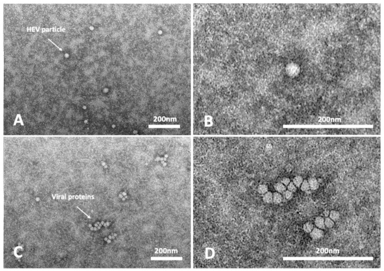
The hepatitis E caused by the virus HEV of genotypes HEV-3 and HEV-4 is a zoonotic foodborne disease spread worldwide. HEV is currently classified into eight different genotypes (HEV-1-8). Genotypes HEV-3 and HEV-4 are zoonotic and are further divided into subtypes. Most of the information on HEV replication remains unknown due to the lack of an efficient cell cultivation system. Over the last couple of years, several protocols for HEV cultivation have been developed on different cell lines; even if they were troublesome, long, and scarcely reproducible, they offered the opportunity to study the replicative cycle of the virus. In the present study, we aimed to obtain a protocol ready to use viral stock in serum free medium that can be used with reduced time of growth and without any purification steps. The employed method allowed isolation and cell adaptation of four swine HEV-3 strains, belonging to three different subtypes. Phylogenetic analyses conducted on partial genome sequences of in vitro isolated strains did not reveal any insertion in the hypervariable region (HVR) of the genomes. A limited number of mutations was acquired in the genome during the virus growth in the partial sequences of Methyltransferase (Met) and ORF2 coding genes.
Keywords: A549; HEV; HEV-3; ORF2; cell culture; isolate; subtype; swine; zoonosis.
Conflict of interest statement
The authors declare no conflict of interest.
Figures


References
-
- Lee G.H., Tan B.H., Teo E.C., Lim S.G., Dan Y.Y., Wee A., Aw P.P., Zhu Y., Hibberd M.L., Tan C.K., et al. Chronic Infection With Camelid Hepatitis E Virus in a Liver Transplant Recipient Who Regularly Consumes Camel Meat and Milk. Gastroenterology. 2016;150:355–357.e3. doi: 10.1053/j.gastro.2015.10.048. - DOI - PubMed
-
- Dalton H.R., van Eijk J.J.J., Cintas P., Madden R.G., Jones C., Webb G.W., Norton B., Pique J., Lutgens S., Devooght-Johnson N., et al. Hepatitis E virus infection and acute non-traumatic neurological injury: A prospective multicentre study. J. Hepatol. 2017;67:925–932. doi: 10.1016/j.jhep.2017.07.010. - DOI - PubMed
Grants and funding
LinkOut - more resources
Full Text Sources
Miscellaneous
