

Potential Therapeutic Strategies for Skeletal Muscle Atrophy
- PMID: 36670909
- PMCID: PMC9854691
- DOI: 10.3390/antiox12010044
Potential Therapeutic Strategies for Skeletal Muscle Atrophy
Abstract
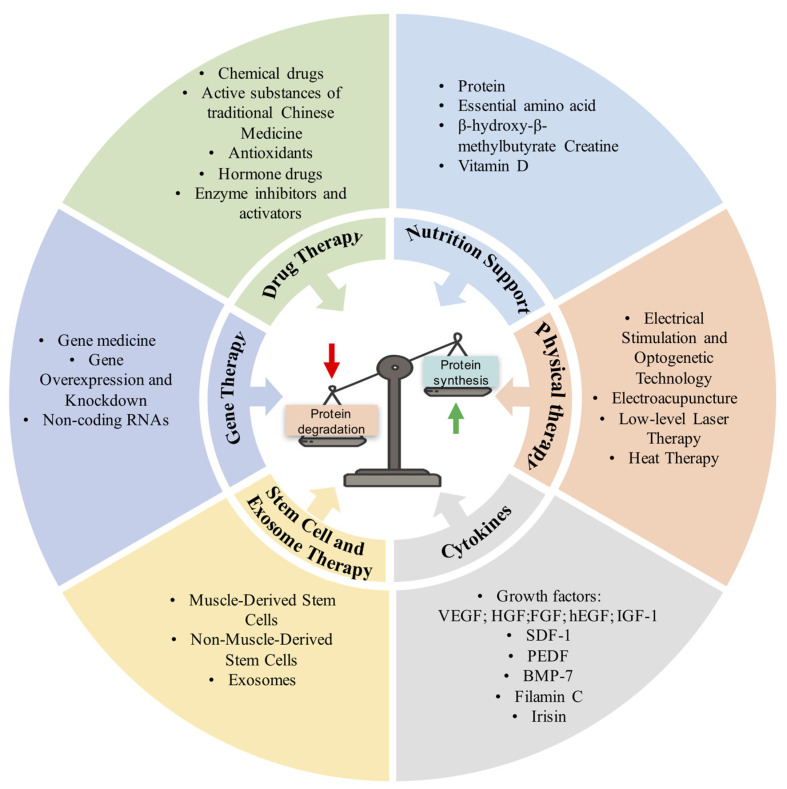
The maintenance of muscle homeostasis is vital for life and health. Skeletal muscle atrophy not only seriously reduces people's quality of life and increases morbidity and mortality, but also causes a huge socioeconomic burden. To date, no effective treatment has been developed for skeletal muscle atrophy owing to an incomplete understanding of its molecular mechanisms. Exercise therapy is the most effective treatment for skeletal muscle atrophy. Unfortunately, it is not suitable for all patients, such as fractured patients and bedridden patients with nerve damage. Therefore, understanding the molecular mechanism of skeletal muscle atrophy is crucial for developing new therapies for skeletal muscle atrophy. In this review, PubMed was systematically screened for articles that appeared in the past 5 years about potential therapeutic strategies for skeletal muscle atrophy. Herein, we summarize the roles of inflammation, oxidative stress, ubiquitin-proteasome system, autophagic-lysosomal pathway, caspases, and calpains in skeletal muscle atrophy and systematically expound the potential drug targets and therapeutic progress against skeletal muscle atrophy. This review focuses on current treatments and strategies for skeletal muscle atrophy, including drug treatment (active substances of traditional Chinese medicine, chemical drugs, antioxidants, enzyme and enzyme inhibitors, hormone drugs, etc.), gene therapy, stem cell and exosome therapy (muscle-derived stem cells, non-myogenic stem cells, and exosomes), cytokine therapy, physical therapy (electroacupuncture, electrical stimulation, optogenetic technology, heat therapy, and low-level laser therapy), nutrition support (protein, essential amino acids, creatine, β-hydroxy-β-methylbutyrate, and vitamin D), and other therapies (biomaterial adjuvant therapy, intestinal microbial regulation, and oxygen supplementation). Considering many treatments have been developed for skeletal muscle atrophy, we propose a combination of proper treatments for individual needs, which may yield better treatment outcomes.
Keywords: cytokine therapy; drug treatment; gene therapy; skeletal muscle atrophy; stem cell therapy.
Conflict of interest statement
The authors declare that the research was conducted in the absence of any commercial or financial relationships that could be construed as a potential conflict of interest.
Figures
References
-
- Qiu J., Fang Q., Xu T., Wu C., Xu L., Wang L., Yang X., Yu S., Zhang Q., Ding F., et al. Mechanistic role of reactive oxygen species and therapeutic potential of antioxidants in denervation- or fasting-induced skeletal muscle atrophy. Front. Physiol. 2018;9:215. doi: 10.3389/fphys.2018.00215. - DOI - PMC - PubMed
Publication types
Grants and funding
LinkOut - more resources
Full Text Sources
Miscellaneous
