Review
. 2023 Jan 12;12(1):177.
doi: 10.3390/antiox12010177.

Lipid Adaptations against Oxidative Challenge in the Healthy Adult Human Brain

Affiliations
Review

Lipid Adaptations against Oxidative Challenge in the Healthy Adult Human Brain

Mariona Jové et al. Antioxidants (Basel). .

Abstract

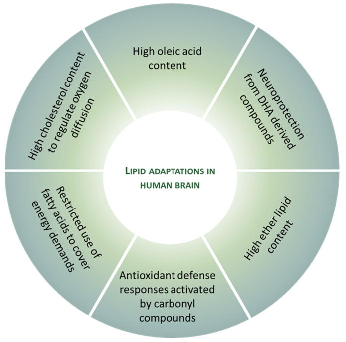
It is assumed that the human brain is especially susceptible to oxidative stress, based on specific traits such as a higher rate of mitochondrial free radical production, a high content in peroxidizable fatty acids, and a low antioxidant defense. However, it is also evident that human neurons, although they are post-mitotic cells, survive throughout an entire lifetime. Therefore, to reduce or avoid the impact of oxidative stress on neuron functionality and survival, they must have evolved several adaptive mechanisms to cope with the deleterious effects of oxidative stress. Several of these antioxidant features are derived from lipid adaptations. At least six lipid adaptations against oxidative challenge in the healthy human brain can be discerned. In this work, we explore the idea that neurons and, by extension, the human brain is endowed with an important arsenal of non-pro-oxidant and antioxidant measures to preserve neuronal function, refuting part of the initial premise.

Keywords: antioxidants; cholesterol; docosahexaenoic acid; fatty acids; lipid peroxidation; lipidomics; oleic acid; plasmalogens.


Conflict of interest statement

The authors declare that the research was conducted in the absence of any commercial or financial relationships that could be construed as a potential conflict of interest.

Figures

Figure 1
Figure 1
Representative lipid categories and specific molecular species present in the adult human brain. Fatty acids (FAs) shown characterize around 80–90% of total FA profile of the human brain ([13,14]). Glycerolipids (GL) in human brain comprise mono-, di-, and tri-acylglycerols. Glycerophospholipids (GP) are the major components ubiquitously found in neural cells; glycerophosphocholines, glycerophosphoethanolamines, glycerophosphoserines, and glycerophosphoinositols are the main GP present in the human brain [8]; molecular species represented are particularly abundant in human brain, highlighting the presence of ether lipids. Sphingolipids (SP) contain a common sphingoid base moiety; they are acylated to form ceramides, which are modified to generate phosphosphingolipids and glycosphingolipids. Sterol lipids comprise cholesterol and its derivatives.
Figure 2
Figure 2
The human brain possesses specific traits at the lipidome level. Among mammalian species, humans included, lipidomes systematically distinguish the brain from the non-neural tissues [47]. This specific lipidome includes an enrichment in ceramides, dihydroceramides, and plasmalogens, and a depletion in fatty amides, triradylglycerols, and sterols. Notably, the extent of differences in the lipidome composition between the brain and non-neural tissues increases in parallel with the increase in the brain function capacity from mice to humans.
Figure 3
Figure 3
Fatty acid profile in the frontal cortex and oleic acid content (%) in different regions of the human brain. (A) Fatty acid distribution in human prefrontal cortex from healthy adult (middle-age) individual. Fatty acid analysis was performed in a gas chromatography system. Data obtained from [14]. (B) Oleic acid content (%) in different regions of the adult human brain. Data obtained from [14]. Brain regions: FC, frontal cortex; FC WM, frontal cortex white matter; FC GM, frontal cortex gray matter; CG, cingulate gyrus; CN, caudate nucleus; V, vermis; EC, entorhinal cortex; H, hippocampus; O, olive; P, putamen; PC, parietal cortex; SN, substantia nigra; T, thalamus; TC, temporal cortex; VC, visual (occipital) cortex.
Figure 4
Figure 4
Lipid adaptations against oxidative challenge in the healthy human brain.

References

    1. Fahy E., Cotter D., Sud M., Subramaniam S. Lipid classification, structures and tools. Biochim. Biophys. Acta-Mol. Cell Biol. Lipids. 2011;1811:637–647. doi: 10.1016/j.bbalip.2011.06.009. - DOI - PMC - PubMed
    1. Yetukuri L., Ekroos K., Vidal-Puig A., Orešič M. Informatics and computational strategies for the study of lipids. Mol. Biosyst. 2008;4:121–127. doi: 10.1039/B715468B. - DOI - PubMed
    1. Han X. Neurolipidomics: Challenges and developments. Front. Biosci. 2007;12:2601. doi: 10.2741/2258. - DOI - PMC - PubMed
    1. Han X., Gross R.W. The foundations and development of lipidomics. J. Lipid Res. 2022;63:100164. doi: 10.1016/j.jlr.2021.100164. - DOI - PMC - PubMed
    1. Fahy E., Subramaniam S., Murphy R.C., Nishijima M., Raetz C.R.H., Shimizu T., Spener F., van Meer G., Wakelam M.J.O., Dennis E.A. Update of the LIPID MAPS comprehensive classification system for lipids. J. Lipid Res. 2009;50:S9–S14. doi: 10.1194/jlr.R800095-JLR200. - DOI - PMC - PubMed