. 2022 Dec 22;13(1):23.
doi: 10.3390/biom13010023.

HIF1A Knockout by Biallelic and Selection-Free CRISPR Gene Editing in Human Primary Endothelial Cells with Ribonucleoprotein Complexes

Affiliations

HIF1A Knockout by Biallelic and Selection-Free CRISPR Gene Editing in Human Primary Endothelial Cells with Ribonucleoprotein Complexes

Camilla Blunk Brandt et al. Biomolecules. .

Abstract

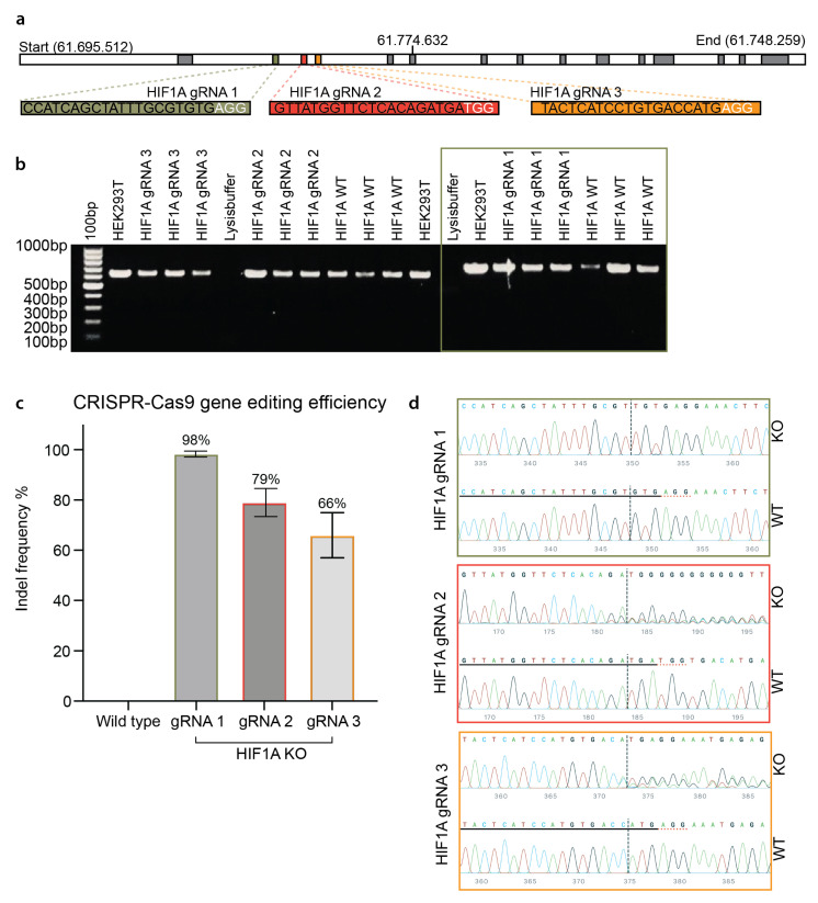
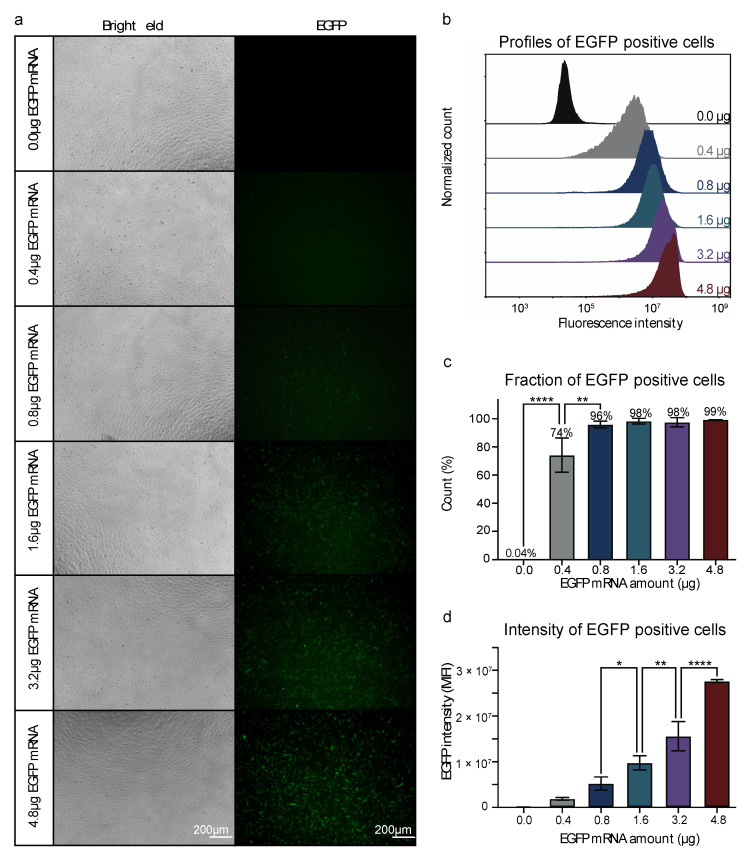
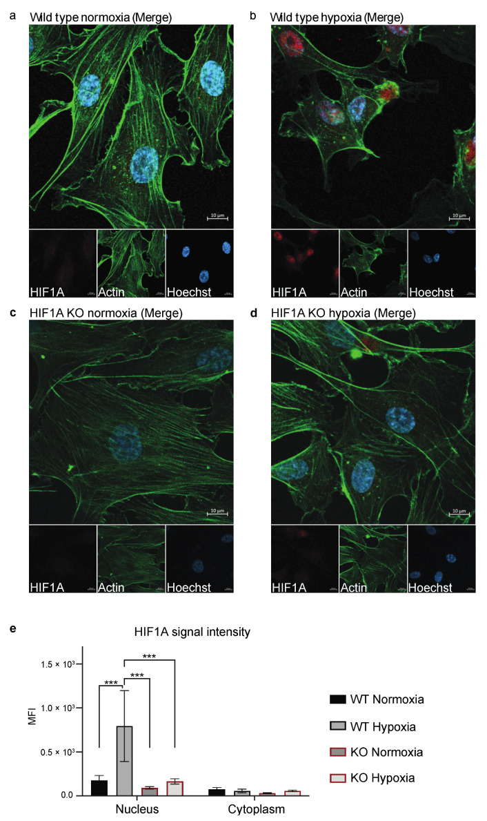
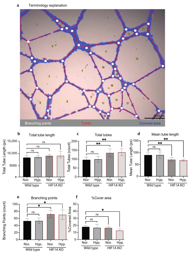
Primary endothelial cells (ECs), especially human umbilical vein endothelial cells (HUVECs), are broadly used in vascular biology. Gene editing of primary endothelial cells is known to be challenging, due to the low DNA transfection efficiency and the limited proliferation capacity of ECs. We report the establishment of a highly efficient and selection-free CRISPR gene editing approach for primary endothelial cells (HUVECs) with ribonucleoprotein (RNP) complex. We first optimized an efficient and cost-effective protocol for messenger RNA (mRNA) delivery into primary HUVECs by nucleofection. Nearly 100% transfection efficiency of HUVECs was achieved with EGFP mRNA. Using this optimized DNA-free approach, we tested RNP-mediated CRISPR gene editing of primary HUVECs with three different gRNAs targeting the HIF1A gene. We achieved highly efficient (98%) and biallelic HIF1A knockout in HUVECs without selection. The effects of HIF1A knockout on ECs' angiogenic characteristics and response to hypoxia were validated by functional assays. Our work provides a simple method for highly efficient gene editing of primary endothelial cells (HUVECs) in studies and manipulations of ECs functions.

Keywords: CRISPR-Cas; HIF1A; endothelial cells; gene editing; human umbilical vein endothelial cells; hypoxia inducible factor 1 alpha; non-viral gene editing; nucleofection; ribonucleoprotein; transfection.


Conflict of interest statement

The authors declare no conflict of interest.

Figures

Figure 2
Figure 2
CRISPR gene editing of the HIF1A gene in HUVECs. (a) Graphical illustration of the HIF1A gene, target exons, and gRNAs. The top panel shows the length of the HIF1A gene (chromosome 14 at position 61.695.512–61.748.259). Three isoforms of the gene are known, which contain 14–15 exons (indicated by the grey boxes). The green, red, and yellow, boxes visualize the three target consecutive exons (exon two, −three, and −four). The sequence of HIF1A gRNA 1, −2, and −3 is captured in green, red, and yellow bars. (b) PCR amplification of the CRISPR gene editing of HUVECs. Visualization of the gel electrophoresis of the amplified DNA from HIF1A gRNA 1, −2, and −3 gene edited HUVECs, HIF1A WT (wild type) HUVECs, HEK293T cells, and the lysis buffer. The 100 bp gene marker is shown in the first well. The green frame indicates the samples amplified by the HIF1A F1/R1 primer sets. (c) CRISPR gene editing efficiency of the three different HIF1A gRNAs. Quantification of the gene editing efficiency of the HIF1A gRNA 1, −2, and −3 and the WT. Values are presented as the mean percentage of indels ± STD. (d) Representative visualization of the Sanger sequencing results for each HIF1A gRNA. For each HIF1A gRNA, the WT HUVECs sequence is shown in the bottom row. The gRNA sequence is underlined with black. The PAM sequence is underlined in dotted red. The editing site is marked by the black dotted vertical line. The sequence of the gene-edited part is shown by wave plots. The nucleotides are color coded as G = guanine, C = cytosine, T = thymine, and A = adenine. (bd) Experiments on HUVECs from three biological donors at p. 2–4 (n = 3). HIF1A gRNA 1–3 experiments are performed with one biological donor at p. 4. HIF1A gRNA 1 experiments were repeated to achieve three biological replicates.
Figure 1
Figure 1
Nucleofection of HUVECs with a titration of EGFP mRNA. (a) Visualization of the EGFP positive cells by fluorescent- and bright field microscopy. (b) The profiles of EGFP-positive HUVECs at each EGFP mRNA concentration. Values are presented as normalized counts of the fluorescent intensity. (c) Fraction of EGFP-positive HUVECs at each EGFP mRNA concentration. Values are presented as the mean percentage of EGFP-positive cells ± SD. Neighboring EGFP mRNA concentrations are compared by one-way ANOVA with Tukey multiple comparisons test, ** p ≤ 0.01, **** p ≤ 0.0001. (d) Median fluorescence intensity of EGFP-positive cells at each EGFP mRNA concentration. Values are presented as the mean ± SD. Neighboring EGFP mRNA concentrations are compared by one-way ANOVA with Tukey multiple comparisons test, * p ≤ 0.05, ** p ≤ 0.01, **** p ≤ 0.0001. (ad): Experiments on HUVECs from one biological donor at p. 5 (n = 3).
Figure 3
Figure 3
Staining of HIF1A in HIF1A KO and WT HUVECs. (ad) One representative image of the stained HIF1A KO and WT HUVECs, which were stained with Hoechst (blue), anti-actin (green), and anti-HIF1A (red). (a) WT HUVECs cultured in normoxic conditions. (b) WT HUVECs cultured in hypoxic conditions. (c) HIF1A KO HUVECs cultured in normoxic conditions. (d) HIF1A KO HUVECs cultured in hypoxic conditions. (e) Quantification of the HIF1A signal intensity (MFI) in the nucleus and the cytoplasm in WT and KO HUVECs in normoxic and hypoxic conditions, which is compared by two-way ANOVA multiple comparisons test, *** p ≤ 0.001. The experiment was performed with one biological replicate at p. 3 (n = 3) and five images were taken per well. Scale bar 10 µm.
Figure 4
Figure 4
Quantification of the tube formation assay of HIF1A KO and WT HUVECs. (a) One representative bright field image of the TFA HUVECs, visualizing the Wimasis terminology which illustrates the tubes in red, the branching points in white, and the covered area in blue. Scale bar 400 µm. (bf) Quantification of the TFA analysis of HIF1A KO and WT HUVECs cultured in normoxic conditions (Nor.) or 2 h hypoxic conditions (Hyp.). (b) The total tube length calculated in pixels. (c) The total amount of tubes, (count). (d) The mean tube length, (pixels). (e) Shows the total branching points, (count). (f) The percentage of covered area. The mean of each test ± STD is plotted, and one-way ANOVA with Tukey multiple comparisons test compared each sample group to the WT HUVECs cultured in normoxic conditions (ns = no significance, * p ≤ 0.05, and ** p ≤ 0.01). (af): Experiments on HUVECs from one biological donor at final p. 3.

References

    1. Fishman A.P. ENDOTHELIUM: A DISTRIBUTED ORGAN OF DIVERSE CAPABILITIES. Ann. N. Y. Acad. Sci. 1982;401:1–8. doi: 10.1111/j.1749-6632.1982.tb25702.x. - DOI - PubMed
    1. Hassan W. The endothelium and endothelin: Beyond vascular reactivity. Ann. Saudi Med. 2006;26:343–345. doi: 10.5144/0256-4947.2006.343. - DOI - PMC - PubMed
    1. Krüger-Genge A., Blocki A., Franke R.P., Jung F. Vascular Endothelial Cell Biology: An Update. Int. J. Mol. Sci. 2019;20:4411. doi: 10.3390/ijms20184411. - DOI - PMC - PubMed
    1. Carmeliet P. Angiogenesis in health and disease. Nat. Med. 2003;9:653–660. doi: 10.1038/nm0603-653. - DOI - PubMed
    1. Aspelund A., Robciuc M.R., Karaman S., Makinen T., Alitalo K. Lymphatic System in Cardiovascular Medicine. Circ. Res. 2016;118:515–530. doi: 10.1161/CIRCRESAHA.115.306544. - DOI - PubMed

Publication types

MeSH terms

LinkOut - more resources