. 2023 Jan 16;13(1):187.
doi: 10.3390/biom13010187.

Intact Transition Epitope Mapping-Force Differences between Original and Unusual Residues (ITEM-FOUR)

Affiliations

Intact Transition Epitope Mapping-Force Differences between Original and Unusual Residues (ITEM-FOUR)

Claudia Röwer et al. Biomolecules. .

Abstract

Antibody-based point-of-care diagnostics have become indispensable for modern medicine. In-depth analysis of antibody recognition mechanisms is the key to tailoring the accuracy and precision of test results, which themselves are crucial for targeted and personalized therapy. A rapid and robust method is desired by which binding strengths between antigens and antibodies of concern can be fine-mapped with amino acid residue resolution to examine the assumedly serious effects of single amino acid polymorphisms on insufficiencies of antibody-based detection capabilities of, e.g., life-threatening conditions such as myocardial infarction. The experimental ITEM-FOUR approach makes use of modern mass spectrometry instrumentation to investigate intact immune complexes in the gas phase. ITEM-FOUR together with molecular dynamics simulations, enables the determination of the influences of individually exchanged amino acid residues within a defined epitope on an immune complex's binding strength. Wild-type and mutated epitope peptides were ranked according to their experimentally determined dissociation enthalpies relative to each other, thereby revealing which single amino acid polymorphism caused weakened, impaired, and even abolished antibody binding. Investigating a diagnostically relevant human cardiac Troponin I epitope for which seven nonsynonymous single nucleotide polymorphisms are known to exist in the human population tackles a medically relevant but hitherto unsolved problem of current antibody-based point-of-care diagnostics.

Keywords: ITEM-FOUR; immune complex analysis; nanoESI mass spectrometry; personalized genomics; single amino acid polymorphism.


Conflict of interest statement

The authors declare no conflict of interest.

Figures

Figure 1
Figure 1
Nano-ESI mass spectra of peptide 1 (ENREVGDWRKNIDAL)—anti-hcTn I antibody immune complex with increasing collision cell voltage differences (∆CV): (a) 4 V, (b) 16 V, (c) 30 V, and (d) 80 V. Charge states are given for the immune complexes (right ion series), and the inlet in (a) shows a zoom of the 25+ and 26+ ion signals of the antibody (0) and the immune complexes (antibody plus one peptide (1) and antibody plus two peptides (2)). Charge states and m/z values for peptide ion signals are given on the left, and the inlet in (c) shows a zoom of the isotopically resolved doubly protonated peptide ion signal. Antibody fragment ion signals are visible between m/z 1200 and m/z 2300 in (d). Solvent: 200 mM ammonium acetate, pH 6.7.
Figure 2
Figure 2
Courses of normalized educt ion intensities of immune complexes of anti-hcTn I antibody and peptides 1 (black squares), 2 (red circles), and 6 (orange triangles) are shown as functions of collision cell voltage differences (∆CV). Each data point is the mean of at least two independent measurements (see Table S1). Vertical bars indicate standard deviations. The sigmoidal-shaped curves were fitted using a Boltzmann function. The curve parameters are given in Table 2.
Figure 3
Figure 3
Nano-ESI mass spectra of peptide 4 (ENREVGDWLKNIDAL)—anti-hcTn I antibody immune complex with increasing collision cell voltage differences (∆CV): (a) 4 V, (b) 16 V, (c) 30 V, and (d) 80 V. Charge states are given for the immune complexes (right ion series), and the inlet in (a) shows a zoom of the 25+ and 26+ ion signals of the antibody (0) and the immune complexes (antibody plus one peptide (1) and antibody plus two peptides (2)). Charge states and m/z values for peptide ion signals are given on the left, and the inlet in (c) shows a zoom of the isotopically resolved doubly protonated peptide ion signal. Antibody fragment ion signals are visible between m/z 1200 and m/z 2300 in (d). Solvent: 200 mM ammonium acetate, pH 6.7.
Figure 4
Figure 4
Courses of normalized educt ion intensities of immune complexes of anti-hcTn I antibody and peptides 3 (blue triangles), 4 (green triangles), 5 (purple squares), and 7 (light blue triangles) are shown as functions of collision cell voltage differences (∆CV). Each data point is the mean of at least two independent measurements (see Table S1). Vertical bars indicate standard deviations. The sigmoidal-shaped curves were fitted using a Boltzmann function. The curve parameters are given in Table 2.
Figure 5
Figure 5
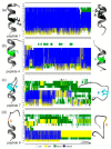
Peptide structure models and secondary structure elements prior to and after molecular dynamics simulations Structure models predicted for epitope peptides (left) were compared to structure models after 50 ns simulations (right). (a) peptide 1, (b) peptide 4, (c) peptide 7, and (d) peptide 6. Amino acid residues of peptides are listed from top to bottom (center). The vertical line at the left indicates the epitope region. The secondary structure element in which each residue is involved at a given simulation time point is depicted from left to right as a color-coded bar (10,000 bars per line). Color code: white: coil; yellow: turn; green: bend; blue: helical.

References

    1. Badrick T. Evidence-based laboratory medicine. Clin. Biochem. Rev. 2013;34:43–46. - PMC - PubMed
    1. Vogenberg F.R., Isaacson Barash C., Pursel M. Personalized medicine: Part 1: Evolution and development into theranostics. Pharm. Ther. 2010;35:560–576. - PMC - PubMed
    1. Reichlin T., Hochholzer W., Bassetti S., Steuer S., Stelzig C., Hartwiger S., Biedert S., Schaub N., Buerge C., Potocki M., et al. Early diagnosis of myocardial infarction with sensitive cardiac troponin assays. N. Engl. J. Med. 2009;361:858–867. doi: 10.1056/NEJMoa0900428. - DOI - PubMed
    1. Keller T., Zeller T., Peetz D., Tzikas S., Roth A., Czyz E., Bickel C., Baldus S., Warnholtz A., Frohlich M., et al. Sensitive troponin I assay in early diagnosis of acute myocardial infarction. N. Engl. J. Med. 2009;361:868–877. doi: 10.1056/NEJMoa0903515. - DOI - PubMed
    1. Ma H., Cassedy A., O’Kennedy R. The role of antibody-based troponin detection in cardiovascular disease: A critical assessment. J. Immunol. Methods. 2021;497:113108. doi: 10.1016/j.jim.2021.113108. - DOI - PMC - PubMed

Publication types