Review
. 2023 Jan 10;12(1):110.
doi: 10.3390/biology12010110.

Extracellular Vesicles: New Classification and Tumor Immunosuppression

Affiliations
Review

Extracellular Vesicles: New Classification and Tumor Immunosuppression

Mona Sheta et al. Biology (Basel). .

Abstract

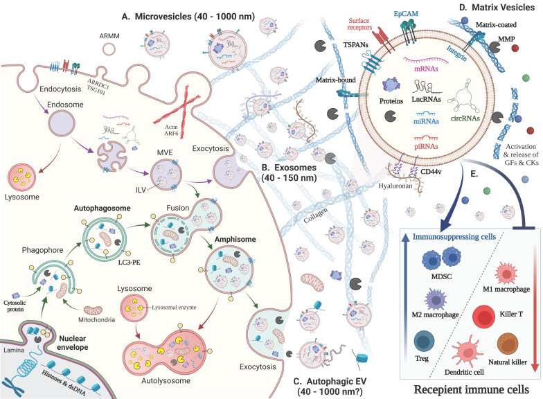
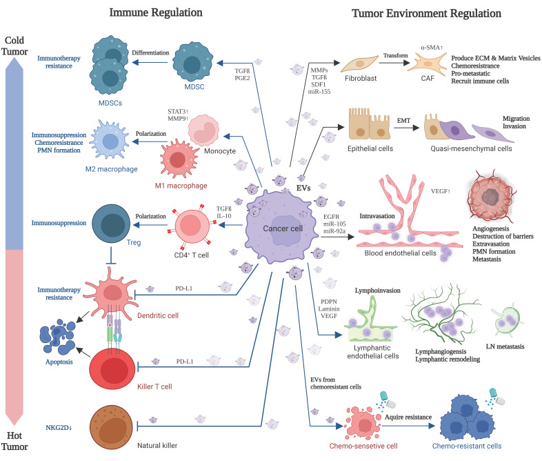
Extracellular vesicles (EVs) are cell-derived membrane-surrounded vesicles carrying various types of molecules. These EV cargoes are often used as pathophysiological biomarkers and delivered to recipient cells whose fates are often altered in local and distant tissues. Classical EVs are exosomes, microvesicles, and apoptotic bodies, while recent studies discovered autophagic EVs, stressed EVs, and matrix vesicles. Here, we classify classical and new EVs and non-EV nanoparticles. We also review EVs-mediated intercellular communication between cancer cells and various types of tumor-associated cells, such as cancer-associated fibroblasts, adipocytes, blood vessels, lymphatic vessels, and immune cells. Of note, cancer EVs play crucial roles in immunosuppression, immune evasion, and immunotherapy resistance. Thus, cancer EVs change hot tumors into cold ones. Moreover, cancer EVs affect nonimmune cells to promote cellular transformation, including epithelial-to-mesenchymal transition (EMT), chemoresistance, tumor matrix production, destruction of biological barriers, angiogenesis, lymphangiogenesis, and metastatic niche formation.

Keywords: amphisome; autophagy; cellular communication; exosome; extracellular vesicle; immune evasion; immunosuppression; matrix vesicle; therapy resistance; tumor microenvironment.


Conflict of interest statement

The authors declare no conflict of interest.

Figures

Figure 1
Figure 1
Biogenesis of various EV types and their immunoregulatory roles. (Left) Biogenesis of various EV types. (A) Microvesicles (40–1000 nm) arise from the outward budding and shedding of the plasma membrane. Small microvesicles called ARMM were recently discovered. (B) Exosomes (40–150 nm) are secreted via exocytosis of intraluminal vesicles (ILVs) by plasma membrane fusion with multivesicular endosomes (MVE) with the plasma membrane (purple arrow). (C) Autophagosome fusion with MVEs generates ‘amphisomes’, whose fusion with the plasma membrane secretes autophagic EVs and vesicle-free cytosolic and nuclear molecules, such as histones and dsDNA (green arrows). This pathway is called ‘exophagy’, a hybrid of exosomes and autophagy. Mitochondrial autophagy is called ‘mitophagy,’ which may result in the secretion of mitochondria. (Upper right) EVs contain a lipid bilayer membrane that protects encapsulated materials, such as proteins, nucleic acids, lipids, and metabolites. The EV surface contains membrane-bound and transmembrane proteins, such as tetraspanins (TSPANs: CD63, CD9, CD81, etc.) and integrins, which bind to matrix proteins. (D) ‘Matrix vesicle’ is a general term for matrix-bound vesicles and matrix-coated vesicles. MMPs cleave matrix components, such as collagen, to release EVs, growth factors (GFs), and chemokines (CKs). (E) Immunoregulatory roles of cancer cell-derived EVs. (i) The suppression and apoptosis of killer T cells, natural killer cells, and dendritic cells; (ii) activation of immunosuppressing cells, such as MDSCs and Tregs; and (iii) polarization of macrophages from the M1 to M2 type.
Figure 2
Figure 2
Cancer cell-derived EVs affect various cell types in the tumor microenvironment. Cancer EVs play crucial roles in immunosuppression, immune evasion, apoptosis of immune cells, and immunotherapy resistance. Thus, cancer EVs change hot tumors into cold ones. Moreover, cancer EVs affect nonimmune cells, including cellular transformation (including EMT), chemoresistance, tumor matrix production, destruction of biological barriers, angiogenesis, lymphangiogenesis, and pro-/premetastatic niche (PMN) formation.

References

    1. Théry C., Witwer K.W., Aikawa E., Alcaraz M.J., Anderson J.D., Andriantsitohaina R., Antoniou A., Arab T., Archer F., Atkin-Smith G.K., et al. Minimal information for studies of extracellular vesicles 2018 (MISEV2018): A position statement of the International Society for Extracellular Vesicles and update of the MISEV2014 guidelines. J. Extracell. Vesicles. 2018;7:1535750. doi: 10.1080/20013078.2018.1535750. - DOI - PMC - PubMed
    1. Jeppesen D.K., Fenix A.M., Franklin J.L., Higginbotham J.N., Zhang Q., Zimmerman L.J., Liebler D.C., Ping J., Liu Q., Evans R., et al. Reassessment of Exosome Composition. Cell. 2019;177:428–445.e18. doi: 10.1016/j.cell.2019.02.029. - DOI - PMC - PubMed
    1. Eguchi T., Sogawa C., Ono K., Matsumoto M., Tran M.T., Okusha Y., Lang B.J., Okamoto K., Calderwood S.K. Cell Stress Induced Stressome Release Including Damaged Membrane Vesicles and Extracellular HSP90 by Prostate Cancer Cells. Cells. 2020;9:755. doi: 10.3390/cells9030755. - DOI - PMC - PubMed
    1. Hikita T., Kuwahara A., Watanabe R., Miyata M., Oneyama C. Src in endosomal membranes promotes exosome secretion and tumor progression. Sci. Rep. 2019;9:3265. doi: 10.1038/s41598-019-39882-z. - DOI - PMC - PubMed
    1. Russell R.C., Tian Y., Yuan H., Park H.W., Chang Y.Y., Kim J., Kim H., Neufeld T.P., Dillin A., Guan K.L. ULK1 induces autophagy by phosphorylating Beclin-1 and activating VPS34 lipid kinase. Nat. Cell Biol. 2013;15:741–750. doi: 10.1038/ncb2757. - DOI - PMC - PubMed

LinkOut - more resources