

A Method for Using Video Presentation to Increase Cortical Region Activity during Motor Imagery Tasks in Stroke Patients
- PMID: 36672012
- PMCID: PMC9855988
- DOI: 10.3390/brainsci13010029
A Method for Using Video Presentation to Increase Cortical Region Activity during Motor Imagery Tasks in Stroke Patients
Abstract
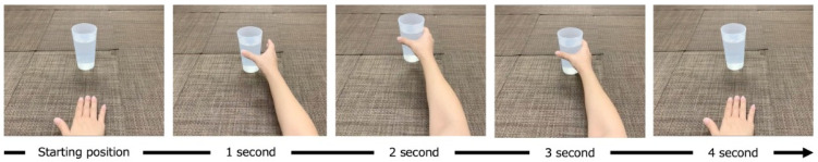
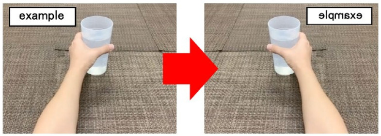
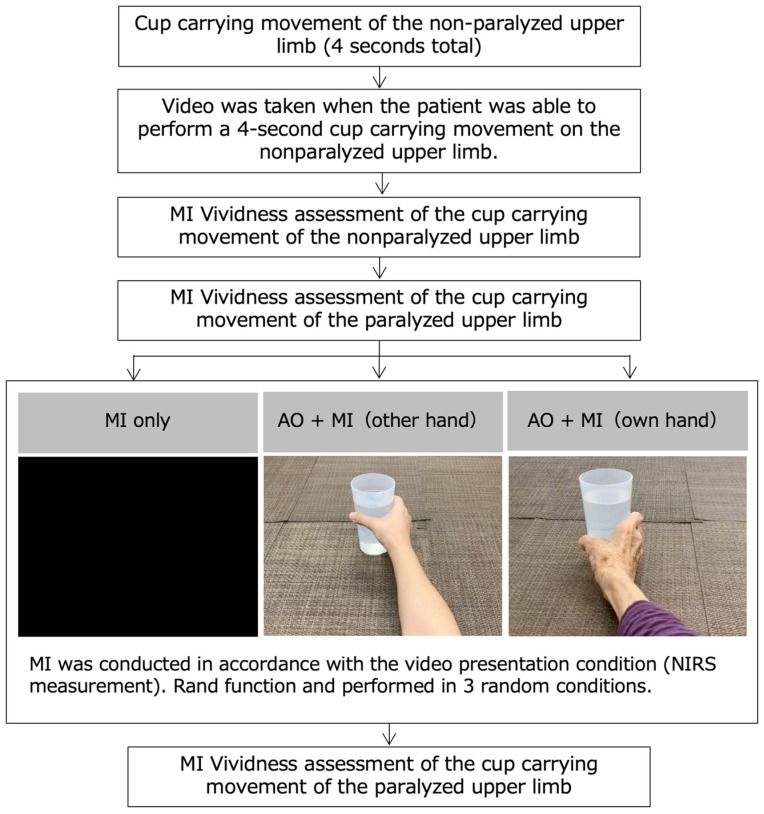
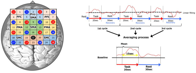
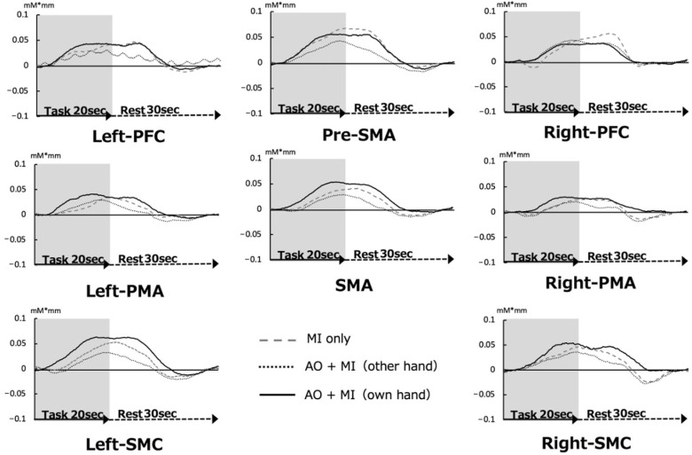
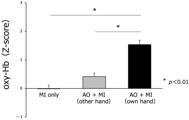
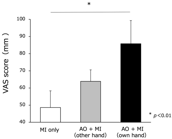
Previous studies have reported that stroke patients have difficulty recalling the motor imagery (MI) of a task, also known as MI vividness. Research on combining MI with action observation is gaining importance as a method to improve MI vividness. We enrolled 10 right-handed stroke patients and compared MI vividness and cortical activity under different presentation methods (no inverted image, inverted image of another individual’s hand, and an inverted image of the patient’s nonparalyzed hand) using near-infrared spectroscopy. Images of the nonparalyzed upper limb were inverted to make the paralyzed upper limb appear as if it were moving. Three tasks (non inverted image, AO + MI (other hand), AO + MI (own hand)) were randomly performed on 10 stroke patients. MI vividness was significantly higher when the inverted image of the nonparalyzed upper limb was presented compared to the other conditions (p < 0.01). The activity of the cortical regions was also significantly enhanced (p < 0.01). Our study highlights the potential application of inverted images of a stroke patient’s own nonparalyzed hand in mental practice to promote the motor recovery of stroke patients. This technique achieved higher levels of MI vividness and cortical activity when performing motor tasks.
Keywords: inverse video; motor imagery vividness; near-infrared spectroscopy; stroke.
Conflict of interest statement
The authors declare no conflict of interest.
Figures
Similar articles
-
Differences in Cortical Area Activity and Motor Imagery Vivid-Ness during Evaluation of Motor Imagery Tasks in Right and Left Hemiplegics.Brain Sci. 2023 Apr 29;13(5):748. doi: 10.3390/brainsci13050748. Brain Sci. 2023. PMID: 37239220 Free PMC article.
-
A method for using video presentation to increase the vividness and activity of cortical regions during motor imagery tasks.Neural Regen Res. 2021 Dec;16(12):2431-2437. doi: 10.4103/1673-5374.313058. Neural Regen Res. 2021. PMID: 33907031 Free PMC article.
-
Enhancing upper-limb neurorehabilitation in chronic stroke survivors using combined action observation and motor imagery therapy.Front Neurol. 2023 Mar 2;14:1097422. doi: 10.3389/fneur.2023.1097422. eCollection 2023. Front Neurol. 2023. PMID: 36937513 Free PMC article.
-
Multiple roles of motor imagery during action observation.Front Hum Neurosci. 2013 Nov 25;7:807. doi: 10.3389/fnhum.2013.00807. Front Hum Neurosci. 2013. PMID: 24324428 Free PMC article. Review.
-
Efficacy of motor imagery additional to motor-based therapy in the recovery of motor function of the upper limb in post-stroke individuals: a systematic review.Top Stroke Rehabil. 2019 Oct;26(7):548-553. doi: 10.1080/10749357.2019.1627716. Epub 2019 Jul 2. Top Stroke Rehabil. 2019. PMID: 31264520
Cited by
-
Combined action observation and motor imagery practice for upper limb recovery following stroke: a systematic review and meta-analysis.Front Neurol. 2025 Jul 23;16:1567421. doi: 10.3389/fneur.2025.1567421. eCollection 2025. Front Neurol. 2025. PMID: 40771975 Free PMC article.
-
Differences in Cortical Area Activity and Motor Imagery Vivid-Ness during Evaluation of Motor Imagery Tasks in Right and Left Hemiplegics.Brain Sci. 2023 Apr 29;13(5):748. doi: 10.3390/brainsci13050748. Brain Sci. 2023. PMID: 37239220 Free PMC article.
References
-
- Buchignani B., Beani E., Pomeroy V., Iacono O., Sicola E., Perazza S., Bieber E., Feys H., Klingels K., Cioni G., et al. Action observation training for rehabilitation in brain injuries: A systematic review and meta-analysis. BMC Neurol. 2019;19:344. doi: 10.1186/s12883-019-1533-x. - DOI - PMC - PubMed
LinkOut - more resources
Full Text Sources
