. 2022 Dec 30;13(1):73.
doi: 10.3390/brainsci13010073.

Hemispherotomy in Infants with Hemimegalencephaly: Long-Term Seizure and Developmental Outcome in Early Treated Patients

Affiliations

Hemispherotomy in Infants with Hemimegalencephaly: Long-Term Seizure and Developmental Outcome in Early Treated Patients

Chiara Pepi et al. Brain Sci. .

Abstract

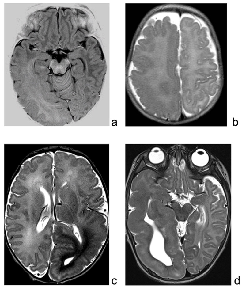
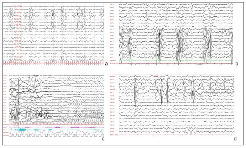
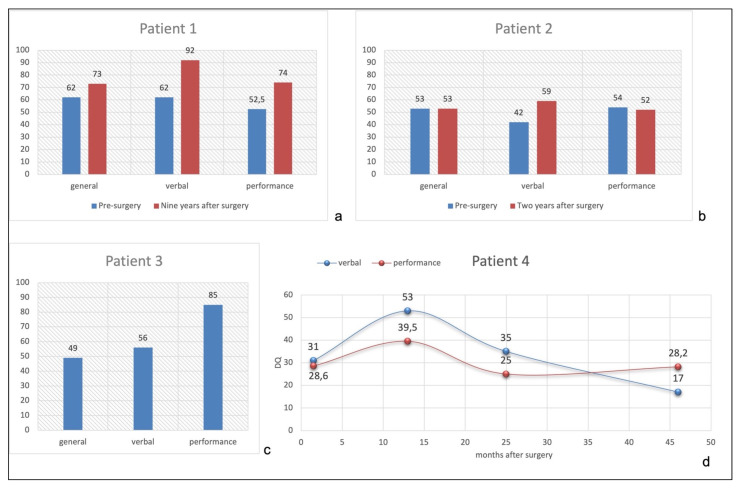
Hemimegalencephaly (HME) is a rare brain congenital malformation, consisting in altered neuronal migration and proliferation within one hemisphere, which is responsible for early onset drug-resistant epilepsy. Hemispherotomy is an effective treatment option for patients with HME and drug-resistant epilepsy. Surgical outcome may be variable among different surgical series, and the long-term neuropsychological trajectory has been rarely defined using a standardized neurocognitive test. We report the epileptological and neuropsychological long-term outcomes of four consecutive HME patients, operated on before the age of three years. All patients were seizure-free and drug-free, and the minimum follow-up duration was of five years. Despite the excellent post-surgical seizure outcome, the long-term developmental outcome is quite variable between patients, ranging from mild to severe intellectual disabilities. Patients showed improvement mainly in communication skills, while visuo-perceptive and coordination abilities were more impaired. Epileptological outcome seems to be improved in early treated patients; however, neuropsychological outcome in HME patients may be highly variable despite early surgery.

Keywords: developmental outcome; epilepsy surgery; hemimegalencephaly; hemispherotomy.


Conflict of interest statement

The authors declare no conflict of interest.

Figures

Figure 1
Figure 1
(a): MRI of patient 1, showing enlarged right hemisphere, with gray–white matter blurring mainly in the temporo-occipital regions. (b): T2-weighted sequence of patient 2 MRI, showing right enlargement of temporo-occipital regions, with blurring, extending also over the suprasylvian regions. (c): MRI of patient 3, showing enlarged and malformed cortex over the left hemisphere, mainly posteriorly. (d): MRI of patient 4, T2-weighted sequence, showing enlargement and malformation of left hemisphere, mainly temporal region.
Figure 2
Figure 2
(a): Interictal EEG of patient 1, showing sub-continuous poly-sharp waves and slow waves with some flattening over the right posterior perisylvian region. (b): Interictal EEG of patient 3, showing pseudo-periodic paroxystic pattern involving the left hemisphere, characterized by repetitive polyspikes. (c): Patient 3. Focal seizure, characterized by rhythmic theta activity involving the left hemisphere and the vertex, without contralateral diffusion. (d): EEG of patient 4, showing continuous and pseudo-periodic spike–wave complexes over the right centro-temporal derivations, with focal seizure starting from central and temporal regions.
Figure 3
Figure 3
(a): Patient 1 pre- and post-surgical quotients. (b): Patient 2 pre- and post-surgical quotients. (c): Patient 3 post-surgical quotients. (d): Patient 4 post-surgical verbal and performance quotients performed at 3.5, 15, 27, and 48 months after surgery.

References

    1. Barkovich A.J., Guerrini R., Kuzniecky R.I., Jackson G.D., Dobyns W.B. A developmental and genetic classification for malformations of cortical development: Update 2012. Brain. 2012;135:1348–1369. doi: 10.1093/brain/aws019. - DOI - PMC - PubMed
    1. Woo C.L.F., Chuang S.H., Becker L.E., Jay V., Otsubo H., Rutka J.T., Snead O. Radiologic-pathologic correlation in focal cortical dysplasia and hemimegalencephaly in 18 children. Pediatr. Neurol. 2001;25:295–303. doi: 10.1016/S0887-8994(01)00318-6. - DOI - PubMed
    1. Baek S.T., Gibbs E.M., Gleeson J.G., Mathern G.W. Hemimegalencephaly, a paradigm for somatic postzygotic neurodevelopmental disorders. Curr. Opin. Neurol. 2013;26:122–127. doi: 10.1097/WCO.0b013e32835ef373. - DOI - PubMed
    1. Di Rocco C., Battaglia D.I., Pietrini D., Piastra M., Massimi L. Hemimegalencephaly: Clinical implications and surgical treatment. Child’s Nerv. Syst. 2006;22:852–866. doi: 10.1007/s00381-006-0149-9. - DOI - PubMed
    1. Garcia C.A.B., Carvalho S.C.S., Yang X., Ball L.L., George R.D., James K.N., Stanley V., Breuss M.W., Thomé U., Santos M.V., et al. mTOR pathway somatic variants and the molecular pathogenesis of hemimegalencephaly. Epilepsia Open. 2020;5:97–106. doi: 10.1002/epi4.12377. - DOI - PMC - PubMed

LinkOut - more resources