. 2023 Jan 7;12(2):257.
doi: 10.3390/cells12020257.

Evaluation of Endocan as a Treatment for Acute Inflammatory Respiratory Failure

Affiliations

Evaluation of Endocan as a Treatment for Acute Inflammatory Respiratory Failure

Maxence Hureau et al. Cells. .

Abstract

Background: Acute respiratory distress syndrome (ARDS) is a life-threatening condition resulting from acute pulmonary inflammation. However, no specific treatment for ARDS has yet been developed. Previous findings suggest that lung injuries related to ARDS could be regulated by endocan (Esm-1). The aim of this study was to evaluate the potential efficiency of endocan in the treatment of ARDS.

Methods: We first compared the features of acute pulmonary inflammation and the severity of hypoxemia in a tracheal LPS-induced acute lung injury (ALI) model performed in knockout (Esm1-/-) and wild type (WT) littermate C57Bl/6 mice. Next, we assessed the effects of a continuous infusion of glycosylated murine endocan in our ALI model in Esm1-/- mice.

Results: In our ALI model, we report higher alveolar leukocytes (p < 0.001), neutrophils (p < 0.001), and MPO (p < 0.001), and lower blood oxygenation (p < 0.001) in Esm1-/- mice compared to WT mice. Continuous delivery of glycosylated murine endocan after LPS-induced ALI resulted in decreased alveolar leukocytes (p = 0.012) and neutrophils (p = 0.012), higher blood oxygenation levels (p < 0.001), and reduced histological lung injury (p = 0.04), compared to mice treated with PBS.

Conclusions: Endocan appears to be an effective treatment in an ARDS-like model in C57Bl/6 mice.

Keywords: Esm-1; acute lung injury; acute respiratory distress syndrome; endocan; respiratory failure.


Conflict of interest statement

L.P. is a former employee of Biothelis. P.L. is the founder of Biothelis. N.D.F.C is a current employee of Biothelis. The funders had no role in the design of the study; in the collection, analyses, or interpretation of data; in the writing of the manuscript; or in the decision to publish the results.

Figures

Figure 1
Figure 1
Experimental protocols. The first protocol (A) was performed to study differences of the LPS induced ALI between WT littermate and Esm1−/− C57Bl/6 mice. The intra tracheal instillation of LPS was performed at D0, the clinical examination, with noninvasive measurement of oximetry, was performed each day, and the mice were sacrificed at different times to describe the features of ALI at each timing. The second protocol (B) was performed to study the effect of the continuous infusion of mouse endocan by osmotic pumps in Esm1−/− mice. Osmotic pumps were filled with either endocan or PBS and implanted subcutaneously one day before i.t. LPS instillation. Clinical examination, including noninvasive measurement of oximetry, was performed daily, and the mice were sacrificed at day 5 to describe the features of ALI. LPS: lipopolysaccharides; i.t.: intra tracheal instillation.
Figure 2
Figure 2
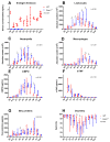
Endogenous endocan alleviates the inflammatory response and improves pulmonary physiological function. The inflammatory response in WT (red) and Esm1−/− (blue) mice after the intratracheal instillation of LPS at different time points. The evolution of endogenous endocan serum concentrations at different timings are shown in (A). The BAL examinations are shown, with the absolute number of leukocytes (B), neutrophils (C), and macrophages (D). mMPO BAL concentrations are shown in (E), mTNF BAL concentrations are shown in (F), and protein BAL concentrations are shown in (G). The evolution of transcutaneous oximetry at different timings is shown in (H). Data are represented by their median, 25th, and 75th percentiles. ∆ represents the estimation of the effect of the group according to the linear mixed model test. ɸ is the result of the Kruskal–Wallis test. * represents a p < 0.05 as the result of the Mann–Whitney test between the variable of each group at a specific timing. H12: 12 h after tracheal instillation of LPS.
Figure 3
Figure 3
Continuous infusion of endocan reduces the inflammatory response and improves the pulmonary physiological function in Esm1−/− mice. The inflammatory response on day 5 after intratracheal instillation of LPS in Esm1−/− mice treated over 3 days by the continuous infusion of murine endocan (purple) compared to PBS (grey) and WT mice (red). BAL examinations are shown with absolute numbers of leukocytes (A), neutrophils (B), and macrophages (C). mMPO BAL concentrations are shown in (D), mTNF BAL concentrations are shown in (E), and protein BAL concentrations are shown in (F). Transcutaneous oximetry measurements at different times are shown in (G). The serum concentration of endocan at D0 is shown in (H). Data are represented by their median, 25th, and 75th percentile. ∆ is equal to the estimation of the effect of the group according to the linear mixed model test. * represents a p < 0.05 as the result of the Mann–Whitney test between the variables of each group at a specific timing.
Figure 4
Figure 4
Continuous infusion of endocan alleviates the histological features of lung injury in Esm1−/− mice. Histological examination of the lung and assessment with the lung injury severity score (LISS) at day 5 after intratracheal instillation of LPS in Esm1−/− mice treated over 3 days by continuous infusion of PBS vs. murine endocan ((A), 200×; (B), 200×). (A), we present the histological photography of an Esm1−/− mouse treated with a continuous infusion of PBS. We can observe patchy neutrophilic infiltrates, hyalines membranes, and fibrin. (B), the histological photography of an Esm1−/− mouse treated with a continuous infusion of murine endocan is shown. (C), we present the results of LISS between the WT (red) and the Esm1−/− (blue) groups and between the Esm1−/− group treated either with PBS (grey) or endocan (purple). Data are represented by their median, 25th, and 75th percentile. * represents p < 0.05 as the result of the Mann–Whitney test between the variables of each group.

References

    1. Bellani G., Laffey J.G., Pham T., Fan E., Brochard L., Esteban A., Gattinoni L., van Haren F., Larsson A., McAuley D.F., et al. Epidemiology, Patterns of Care, and Mortality for Patients With Acute Respiratory Distress Syndrome in Intensive Care Units in 50 Countries. JAMA. 2016;315:788–800. doi: 10.1001/jama.2016.0291. - DOI - PubMed
    1. Ware L.B., Matthay M.A. The Acute Respiratory Distress Syndrome. N. Engl. J. Med. 2000;342:1334–1349. doi: 10.1056/NEJM200005043421806. - DOI - PubMed
    1. Ley K., Laudanna C., Cybulsky M.I., Nourshargh S. Getting to the Site of Inflammation: The Leukocyte Adhesion Cascade Updated. Nat. Rev. Immunol. 2007;7:678–689. doi: 10.1038/nri2156. - DOI - PubMed
    1. Lassalle P., Molet S., Janin A., Heyden J.V., Tavernier J., Fiers W., Devos R., Tonnel A.B. ESM-1 Is a Novel Human Endothelial Cell-Specific Molecule Expressed in Lung and Regulated by Cytokines. J. Biol. Chem. 1996;271:20458–20464. doi: 10.1074/jbc.271.34.20458. - DOI - PubMed
    1. Gaudet A., Portier L., Prin M., Copin M.-C., Tsicopoulos A., Mathieu D., Lassalle P., De Freitas Caires N. Endocan Regulates Acute Lung Inflammation through Control of Leukocyte Diapedesis. J. Appl. Physiol. 2019;127:668–678. doi: 10.1152/japplphysiol.00337.2019. - DOI - PubMed

Publication types

Substances