

Multipotent Mesenchymal Stromal Cells from Porcine Bone Marrow, Implanted under the Kidney Capsule, form an Ectopic Focus Containing Bone, Hematopoietic Stromal Microenvironment, and Muscles
- PMID: 36672203
- PMCID: PMC9857022
- DOI: 10.3390/cells12020268
Multipotent Mesenchymal Stromal Cells from Porcine Bone Marrow, Implanted under the Kidney Capsule, form an Ectopic Focus Containing Bone, Hematopoietic Stromal Microenvironment, and Muscles
Abstract
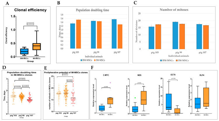
Multipotent mesenchymal stromal cells (MSCs) are an object of intense investigation due to their therapeutic potential. MSCs have been well studied in vitro, while their fate after implantation in vivo has been poorly analyzed. We studied the properties of MSCs from the bone marrow (BM-MSC) before and after implantation under the renal capsule using a mini pig model. Autologous BM-MSCs were implanted under the kidney capsule. After 2.5 months, ectopic foci containing bones, foci of ectopic hematopoiesis, bone marrow stromal cells and muscle cells formed. Small pieces of the implant were cultivated as a whole. The cells that migrated out from these implants were cultured, cloned, analyzed and were proven to meet the most of criteria for MSCs, therefore, they are designated as MSCs from the implant-IM-MSCs. The IM-MSC population demonstrated high proliferative potential, similar to BM-MSCs. IM-MSC clones did not respond to adipogenic differentiation inductors: 33% of clones did not differentiate, and 67% differentiated toward an osteogenic lineage. The BM-MSCs revealed functional heterogeneity after implantation under the renal capsule. The BM-MSC population consists of mesenchymal precursor cells of various degrees of differentiation, including stem cells. These newly discovered properties of mini pig BM-MSCs reveal new possibilities in terms of their manipulation.
Keywords: differentiation; ectopic foci; implantation; multipotent mesenchymal stromal cells (MSCs); proliferation.
Conflict of interest statement
The authors declare that the research was conducted in the absence of any commercial or financial relationships that could be construed as a potential conflict of interest.
Figures


Similar articles
-
Single-cell RNA sequencing deconvolutes the in vivo heterogeneity of human bone marrow-derived mesenchymal stem cells.Int J Biol Sci. 2021 Oct 11;17(15):4192-4206. doi: 10.7150/ijbs.61950. eCollection 2021. Int J Biol Sci. 2021. PMID: 34803492 Free PMC article.
-
Periosteal skeletal stem cells can migrate into the bone marrow and support hematopoiesis after injury.Elife. 2025 May 22;13:RP101714. doi: 10.7554/eLife.101714. Elife. 2025. PMID: 40401637 Free PMC article.
-
Evaluation of Porcine Versus Human Mesenchymal Stromal Cells From Three Distinct Donor Locations for Cytotherapy.Front Immunol. 2020 May 6;11:826. doi: 10.3389/fimmu.2020.00826. eCollection 2020. Front Immunol. 2020. PMID: 32435248 Free PMC article.
-
Chimerism of bone marrow mesenchymal stem/stromal cells in allogeneic hematopoietic cell transplantation: is it clinically relevant?Chimerism. 2013 Jul-Sep;4(3):78-83. doi: 10.4161/chim.25609. Epub 2013 Jul 11. Chimerism. 2013. PMID: 23880502 Free PMC article. Review.
-
Same or not the same? Comparison of adipose tissue-derived versus bone marrow-derived mesenchymal stem and stromal cells.Stem Cells Dev. 2012 Sep 20;21(14):2724-52. doi: 10.1089/scd.2011.0722. Epub 2012 May 9. Stem Cells Dev. 2012. PMID: 22468918 Review.
Cited by
-
Prospects for the Application of Transplantation With Human Amniotic Membrane Epithelial Stem Cells in Systemic Lupus Erythematosus.Cell Transplant. 2024 Jan-Dec;33:9636897241236586. doi: 10.1177/09636897241236586. Cell Transplant. 2024. PMID: 38469823 Free PMC article. Review.
-
Exploring functional metabolites and proteomics biomarkers in late-preterm and natural-born pigs.Front Vet Sci. 2024 Apr 24;11:1340849. doi: 10.3389/fvets.2024.1340849. eCollection 2024. Front Vet Sci. 2024. PMID: 38721154 Free PMC article.
References
-
- Costa L.A., Eiro N., Fraile M., Gonzalez L.O., Saá J., Garcia-Portabella P., Vega B., Schneider J., Vizoso F.J. Functional heterogeneity of mesenchymal stem cells from natural niches to culture conditions: Implications for further clinical uses. Cell. Mol. Life Sci. 2021;78:447–467. doi: 10.1007/s00018-020-03600-0. - DOI - PMC - PubMed
-
- Sun K., Zhou Z., Ju X., Zhou Y., Lan J., Chen D., Chen H., Liu M., Pang L. Combined transplantation of mesenchymal stem cells and endothelial progenitor cells for tissue engineering: A systematic review and meta-analysis. Stem Cell Res. Ther. 2016;7:151. doi: 10.1186/s13287-016-0390-4. - DOI - PMC - PubMed
-
- Shanbhag S., Pandis N., Mustafa K., Nyengaard J.R., Stavropoulos A. Cell Cotransplantation Strategies for Vascularized Craniofacial Bone Tissue Engineering: A Systematic Review and Meta-Analysis of Preclinical In Vivo Studies. Tissue Eng. Part B Rev. 2017;23:101–117. doi: 10.1089/ten.teb.2016.0283. - DOI - PubMed
MeSH terms
LinkOut - more resources
Full Text Sources
