Review
. 2023 Jan 11;12(2):276.
doi: 10.3390/cells12020276.

Macrophages: From Simple Phagocyte to an Integrative Regulatory Cell for Inflammation and Tissue Regeneration-A Review of the Literature

Affiliations
Review

Macrophages: From Simple Phagocyte to an Integrative Regulatory Cell for Inflammation and Tissue Regeneration-A Review of the Literature

Andreas Mamilos et al. Cells. .

Abstract

The understanding of macrophages and their pathophysiological role has dramatically changed within the last decades. Macrophages represent a very interesting cell type with regard to biomaterial-based tissue engineering and regeneration. In this context, macrophages play a crucial role in the biocompatibility and degradation of implanted biomaterials. Furthermore, a better understanding of the functionality of macrophages opens perspectives for potential guidance and modulation to turn inflammation into regeneration. Such knowledge may help to improve not only the biocompatibility of scaffold materials but also the integration, maturation, and preservation of scaffold-cell constructs or induce regeneration. Nowadays, macrophages are classified into two subpopulations, the classically activated macrophages (M1 macrophages) with pro-inflammatory properties and the alternatively activated macrophages (M2 macrophages) with anti-inflammatory properties. The present narrative review gives an overview of the different functions of macrophages and summarizes the recent state of knowledge regarding different types of macrophages and their functions, with special emphasis on tissue engineering and tissue regeneration.

Keywords: M1-macrophages; M2-macrophages; biomaterials; inflammation; macrophage; monocytes; plasticity; tissue regeneration.


Conflict of interest statement

The authors declare no conflict of interest.

Figures

Figure 1
Figure 1
Formation and differentiation of macrophages. Schematic depiction of the differentiation of monocytes during embryonic development unfolding in three sequential waves and adulthood as well as their further differentiation to different subpopulations of macrophages in the tissue [25] (AGM region: aorta-gonad-mesonephros region; EMP: Erythro-Myeloid Progenitors, YS: yolk sac), modified after Corliss et al. [26].
Figure 2
Figure 2
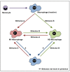
Plasticity of macrophages: a brief schematic depiction of the plasticity of macrophages from an inactive macrophage (MΦ) into either an M1-macrophage or M2-macrophage according to different stimuli. The scheme represents macrophages as an activations state of cells that can be changed along a continuum into the different sub-populations according to various stimuli in the environment. In addition, M1 and M2 macrophages can be turned into inactivated macrophages if there is a lack of stimuli.
Figure 3
Figure 3
Activation of classically activated macrophages. In the presence of INF-γ-receptor ligands or PAMP, the macrophages adopt the M1 phenotype, which is characterized by the expression of INF-β and TNF-α –receptors. Furthermore, these cells are now capable of synthesizing TNF-α and INF-β and thus achieving self-activation.
Figure 4
Figure 4
Summary of the basic functions of M1-Macrophages.
Figure 5
Figure 5
The activation of M2a-Macrophages. In the presence of IL-4- and IL-13- receptor ligands, the macrophages adopt the M2a phenotype, which is characterized by the expression of INF-β and TNF-α –receptors. Furthermore, these cells are now able to synthesize TNF-α and INF-β and thus undergo self-activation.
Figure 6
Figure 6
Summary of the function of M2a-macrophages at the cellular level.
Figure 7
Figure 7
The polarization of M2b-Macrophages. In the presence of immune complexes, TLR-ligands, and IL-1 receptor ligands, the macrophages adopt the M2b phenotype.
Figure 8
Figure 8
The polarization to M2c-Macrophages. In the presence of glucocorticoids (GC), IL-10 or TGF-β receptor ligands, the macrophages adopt the M2c phenotype, which is capable of synthesizing IL-10 and TGF-β and thus reaching a state of self-activation.

Similar articles

Cited by

References

    1. Gordon S. Elie Metchnikoff, the Man and the Myth. J. Innate Immun. 2016;8:223–227. doi: 10.1159/000443331. - DOI - PMC - PubMed
    1. Xia Z., Triffitt J.T. A review on macrophage responses to biomaterials. Biomed. Mater. 2006;1:R1–R9. doi: 10.1088/1748-6041/1/1/R01. - DOI - PubMed
    1. Erwig L.P., Henson P.M. Clearance of apoptotic cells by phagocytes. Cell Death Differ. 2008;15:243–250. doi: 10.1038/sj.cdd.4402184. - DOI - PubMed
    1. Flannagan R.S., Jaumouillé V., Grinstein S. The cell biology of phagocytosis. Annu. Rev. Pathol. 2012;7:61–98. doi: 10.1146/annurev-pathol-011811-132445. - DOI - PubMed
    1. Braga T.T., Agudelo J.S., Camara N.O. Macrophages During the Fibrotic Process: M2 as Friend and Foe. Front. Immunol. 2015;6:602. doi: 10.3389/fimmu.2015.00602. - DOI - PMC - PubMed

Publication types

Substances

LinkOut - more resources