. 2023 Jan 11;12(2):287.
doi: 10.3390/cells12020287.

SGC-CAMKK2-1: A Chemical Probe for CAMKK2

Affiliations

SGC-CAMKK2-1: A Chemical Probe for CAMKK2

Carrow Wells et al. Cells. .

Abstract

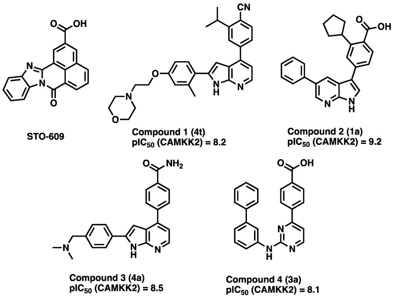
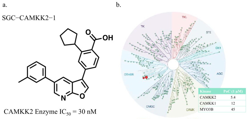
The serine/threonine protein kinase calcium/calmodulin-dependent protein kinase kinase 2 (CAMKK2) plays critical roles in a range of biological processes. Despite its importance, only a handful of inhibitors of CAMKK2 have been disclosed. Having a selective small molecule tool to interrogate this kinase will help demonstrate that CAMKK2 inhibition can be therapeutically beneficial. Herein, we disclose SGC-CAMKK2-1, a selective chemical probe that targets CAMKK2.

Keywords: CAMKK2; NanoBRET; chemical probe; kinase; kinase selectivity.


Conflict of interest statement

D.E.F. has received research funding from GTx, Inc. and has familial relationships with Hummingbird Bioscience, Maia Biotechnology, Alms Therapeutics, Hinova Pharmaceuticals, and Barricade Therapeutics. The funders had no role in the conceptualization of the study or writing of the manuscript, or in the decision to publish this article. The authors declare no conflict of interest.

Figures

Figure 1
Figure 1
STO-609 and some reported (Price et al. numbering noted in parenthesis [47]) CAMKK2 inhibitors. Potencies are depicted as pIC50 values. pIC50 values are the negative log of the IC50 value (in molar). Thus, a pIC50 = 6 corresponds to an IC50 of 1μM, a pIC50 = 7 corresponds to an IC50 = 100 nM, a pIC50 = 8 corresponds to an IC50 = 10 nM, and a pIC50 = 9 corresponds to an IC50 = 1 nM.
Scheme 1
Scheme 1
Synthetic route to compounds 5 and 6. Reagents and conditions (a) 8, Pd(dppf)Cl2 (0.05 eq.), Cs2CO3 (2 eq.), 1,4-dioxane/water (4:1), 120 °C microwave, 30 min; (b) ArB(OH)2 (1 eq.), Pd(dppf)Cl2 (0.05 eq.), Cs2CO3 (2 eq.), 1,4-dioxane/water (4:1), 120 °C microwave, 30 min; (c) 1 N NaOH, MeOH, 75 °C then aq. HCl, 1 h.
Figure 2
Figure 2
(a) The structure and enzyme inhibition IC50 of the CAMKK2 probe molecule. (b) The selectivity of compound 5 (SGC-CAMKK2-1) depicted using a TREEspot diagram obtained from profiling SGC-CAMKK2-1 at 1 µM in the KinomeSCAN® assay panel. PoC = percentage of control. A PoC of 100 indicates no binding and a PoC = 0 is reflective of complete binding. Thus, kinases with PoC values closer to zero are the assay hits and are the kinases to which our compound binds.
Figure 3
Figure 3
Structure and characterization of the negative control compound SGC-CAMKK2-1N (all data n = 1).
Scheme 2
Scheme 2
Synthesis of negative control 7. Reagents and conditions: (a) PhB(OH)2, Cs2CO3, Pd(PPh3)4, DMF/H2O, 16 h; (b) i. CHCl3, Br2, ii. 10% NaOH, MeOH; (c) 4-borono-2-chlorobenzoic acid, Pd(PPh3)4, Na2CO3, dioxane/H2O, 90 °C, 16 h.
Figure 4
Figure 4
Predicted binding mode of probe SGC-CAMKK1 to CAMKK2 kinase domain. (a) Detailed view of the binding interactions between compound 6, a close furopyridine analog of probe SGC-CAMKK-1, and CAMKK2 ATP-binding pocket as seen in the complex co-crystal structure (PDB ID 5UY6). (b) Predicted binding mode of probe SGC-CAMKK1 to CAMKK2 ATP-binding pocket obtained by in silico docking (see Methods for details). In (a,b), dashed black lines indicate potential hydrogen bonds, water molecules are depicted as red spheres, carbon atoms are shown in green (ligand) or yellow (protein), the kinase domain α-helix C is shown in blue, and protein surface (for the bottom of the ATP-binding site) is shown in white.
Figure 5
Figure 5
NanoBRET in-cell target engagement of CAMKK2 probe (5) (n = 3) and STO-609 (n = 1) in HEK293 cells transiently transfected with NLuc-CAMKK2. Increasing doses of inhibitor displaces the fluorescent inhibitor ligand (called a tracer) used in the assay, leading to a reduction in the BRET signal.
Figure 6
Figure 6
Western blot analysis of endogenous CAMKK2 inhibition using p-AMPK(Thr172) levels as a marker of CAMKK2 cellular activity. Western blot images (top) and densitometry analyses (bottom) of C4-2 cells treated with increasing doses of (a) STO-609, (b) probe SGC-CAMKK2-1 (5), and (c) negative control SGC-CAMKK2-1N (7). IC50 for STO-609 = 10.7 μM, SGC-CAMKK2-1 probe = 1.6 μM, and negative control SGC-CAMKK2-1N ≥10 μM. Results represent values from three individual experiments.
Figure 7
Figure 7
The probe compound SGC-CAMKK2-1 (5) inhibits CAMKK2 activity with higher efficacy than STO-609 in MDA-MB 231 triple-negative metastatic breast cancer cells. MDA-MB 231 cells were treated with increasing doses of STO-609 or 5 for 24 h as indicated. Compound 7 (10 μM) was used as the negative control for CAMKK2 inhibition (data not shown). Lysates were collected and immunoblotting was done to check for levels of p-AMPK(Thr172), a substrate of CAMKK2. (a) Representative image showing 5 inhibiting CAMKK2 downstream activity with improved efficacy compared to STO-609. (b) Quantification of immunoblot bands. Results represent values from three individual experiments. * p < 0.05.
Figure 8
Figure 8
Plasma concentration of SGC-CAMKK2-1 and STO-609 in a single dose IP mouse study. The compounds were administered at a dose of 10 mg/kg via IP administration to three animals for each compound. Blood was collected at t = 0.5, 1, 3, and 8 h and immediately spun for 3 min and the plasma was collected, and compound levels were quantified with HPLC/MS.

References

    1. Anderson K.A., Ribar T.J., Lin F., Noeldner P.K., Green M.F., Muehlbauer M.J., Witters L.A., Kemp B.E., Means A.R. Hypothalamic CaMKK2 contributes to the regulation of energy balance. Cell Metab. 2008;7:377–388. doi: 10.1016/j.cmet.2008.02.011. - DOI - PubMed
    1. Pritchard Z.J., Cary R.L., Yang C., Novack D.V., Voor M.J., Sankar U. Inhibition of CaMKK2 reverses age-associated decline in bone mass. Bone. 2015;75:120–127. doi: 10.1016/j.bone.2015.01.021. - DOI - PMC - PubMed
    1. Cary R.L., Waddell S., Racioppi L., Long F., Novack D.V., Voor M.J., Sankar U. Inhibition of Ca(2)(+)/calmodulin-dependent protein kinase kinase 2 stimulates osteoblast formation and inhibits osteoclast differentiation. J. Bone Min. Res. 2013;28:1599–1610. doi: 10.1002/jbmr.1890. - DOI - PMC - PubMed
    1. Williams J.N., Kambrath A.V., Patel R.B., Kang K.S., Mevel E., Li Y., Cheng Y.H., Pucylowski A.J., Hassert M.A., Voor M.J., et al. Inhibition of CaMKK2 Enhances Fracture Healing by Stimulating Indian Hedgehog Signaling and Accelerating Endochondral Ossification. J. Bone Min. Res. 2018;33:930–944. doi: 10.1002/jbmr.3379. - DOI - PMC - PubMed
    1. Broxmeyer H.E., Ropa J., Capitano M.L., Cooper S., Racioppi L., Sankar U. CaMKK2 Knockout Bone Marrow Cells Collected/Processed in Low Oxygen (Physioxia) Suggests CaMKK2 as a Hematopoietic Stem to Progenitor Differentiation Fate Determinant. Stem Cell Rev. Rep. 2022;18:2513–2521. doi: 10.1007/s12015-021-10306-8. - DOI - PMC - PubMed

Publication types

LinkOut - more resources