. 2023 Jan 14;12(2):315.
doi: 10.3390/cells12020315.

Gradient Magnetic Field Accelerates Division of E. coli Nissle 1917

Affiliations

Gradient Magnetic Field Accelerates Division of E. coli Nissle 1917

Svitlana Gorobets et al. Cells. .

Abstract

Cell-cycle progression is regulated by numerous intricate endogenous mechanisms, among which intracellular forces and protein motors are central players. Although it seems unlikely that it is possible to speed up this molecular machinery by applying tiny external forces to the cell, we show that magnetic forcing of magnetosensitive bacteria reduces the duration of the mitotic phase. In such bacteria, the coupling of the cell cycle to the splitting of chains of biogenic magnetic nanoparticles (BMNs) provides a biological realization of such forcing. Using a static gradient magnetic field of a special spatial configuration, in probiotic bacteria E. coli Nissle 1917, we shortened the duration of the mitotic phase and thereby accelerated cell division. Thus, focused magnetic gradient forces exerted on the BMN chains allowed us to intervene in the processes of division and growth of bacteria. The proposed magnetic-based cell division regulation strategy can improve the efficiency of microbial cell factories and medical applications of magnetosensitive bacteria.

Keywords: bacterial division; biomagnetic effects; intracellular forces; magnetic field; mitosis.


Conflict of interest statement

The authors declare no conflict of interest.

Figures

Figure 1
Figure 1
Magnetic system for cultivation of EcN bacteria. In (a,b), 1—permanent magnet, 2—magnetic circuit, and 3—working volume of the magnetic system for cultivation of the EcN. The magnetic field flux density was measured to be a maximum of 0.15 T inside the working volume of the magnetic system. The measurement was carried out by means of a Magnetic Field Flux Density Meter Ш1-8 with Hall-type sensor. The size of the magnetic field sensor (Magnetic Field Flux Density Meter Ш1-8) is 6 mm. Therefore, the specified sensor can measure the magnetic field flux density averaged on the scale of the characteristic dimensions of the sensor at the center of the working volume (3). The red frame shows the unit cell of the magnetic system used for calculation of both the magnetic field flux density distribution and the magnetic field gradient distribution in the working volume of the magnetic system. (c,d) Unit of the magnetic system and the spatial distribution of the MF within it. The magnetization of the magnets is directed along the x-axis. The magnetic field flux density at the central area is 150 mT.
Figure 2
Figure 2
Spatial magnetic field and gradient distributions within a magnetic unit of the bacteria cultivation system: (a) 3D plot of the magnetic field strength modulus, H/M vs. x- and z-coordinates (both in mm); (b) vector field of H vs. x- and z-coordinates in the plane y = 0; (c) vector field of dH/dx vs. the x- and z-coordinates in the plane y = 0; (d) vector field of dH/dz vs. the x- and z-coordinates in the plane y = 0.
Figure 3
Figure 3
Two permanent magnets generate a gradient magnetic field. The magnetic gradient reaches its maximum just above the contact surface (1).
Figure 4
Figure 4
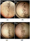
Focusing of EcN bacteria in a gradient MF above the contact surface of the two permanent magnets shown in Figure 3: (a) bacterial cells cultivated on standard medium (control); (b) bacterial cells cultivated on standard medium with the addition of chelates; (c) bacterial cells cultivated on standard medium under the influence of external MF with magnetic field flux density 1500 Oe (0.15 T); (d) bacterial cells cultivated on a standard medium with the addition of chelates under the influence of external MF with magnetic field flux density 1500 Oe (0.15 T).
Figure 5
Figure 5
AFM (a) and MFM (b) images of EcN cells cultivated on standard medium (control). The left image represents AFM of EcN cells, i.e., topography of the surface of EcN cells at the cover glass. The color bar for left image shows the height in nanometers illustrating the shape and size of EcN cells. The right image represents MFM (of the same EcN cells as in left image) in the dynamic MFM mode. The abrupt change of color between inside and outside regions of EcN cells in the right image means that the force of magnetic interaction of the magnetic tip of the cantilever with the EcN cells differs in a step-like manner from the force of interaction with the diamagnetic cover glass.
Figure 6
Figure 6
AFM (a) and MFM (b) images of EcN cells cultivated on medium supplemented with iron chelate. The left image represents the AFM of EcN cells, i.e., the topography of the surface of EcN cells at the cover glass. The color bar for the left image shows the height in micrometers illustrating the shape and size of EcN cells. The right image represents the MFM (of the same EcN cells as in left image) in the dynamic MFM mode. The abrupt change in the colors between the inside and outside regions of EcN cells in the right image means that the force of magnetic interaction of the magnetic tip of the cantilever with the EcN cells differs in a step-like manner from that the force of interaction with the diamagnetic cover glass.
Figure 7
Figure 7
Width and surface area (both normalized to the controls) of the strips, formed by the EcN cell just above the contact surface of the system of two permanent magnets: ST—standard medium (control), ST + MF—standard medium in the external 0.15 T MF, ST + Fe—standard medium with iron chelates, ST + MF + Fe—standard medium with iron chelates in the external 0.15 T MF. The examples of optic images of strips of magnetic focusing of EcN bacteria in a gradient MF above the contact surface of the two permanent magnets are presented in Figure 4. Blue bars represent the mean strip width, while the orange bars are the mean strip area measured using the optic images and the Gwyddion free software [54]. The asterisk (*) means p values less than 0.05.
Figure 8
Figure 8
Distributions of the cell cluster sizes of EcN bacteria: N is the number of bacterial cell clusters, and Nmax is the total number of bacterial cell clusters analyzed. ST—standard medium (control), ST + Fe—standard medium with iron chelates, ST + MF—standard medium in the gradient 0.15 T MF, ST + MF + Fe—standard medium with iron chelates in the gradient MF of 0.15 T. The x-axis refers to the size of bacterial cell clusters in the suspension used to study magnetophoretic velocity.
Figure 9
Figure 9
Calculated spatial distribution of ∇B above the magnets. The blue arrows show the gradient directions, which are the same as the magnetic gradient force directions, while the white arrows represent the magnetization of BMNs in bacteria.
Figure 10
Figure 10
Magnetic field distributions and bacterial division. (a) Vector field of the magnetic field strength, H (the blue arrows). (b) Spatial distribution of its modulus above the two magnets of the lateral half size; a1 = 23 mm and thickness 2a3 = 10 mm. The field was calculated in the plane 0.2 mm above the top of the magnets. The white arrows show the directions of the magnetizations of the magnets. The legend shows the values of |H|/M. (c) Bacterial division by the magnetic gradient forces and the magnetic field distribution (blue arrows); the red arrows are the x-components of the forces acting on the BMN chains in the magnetic system shown in Figure 10. (d) Vector field of the magnetic gradient in unit of the cell incubator shown in Figure 1 (the horizontal and vertical axes are the x- and z-coordinates, respectively).
Figure 11
Figure 11
Calculated magnetic field strength components and their derivatives: (a) Hz/M, (b) Hx/M, (c) (dHz/dx)M−1, and (d) (dHx/dx)M−1 (both derivatives are in mm−1), versus the x- coordinate (in mm) The calculations were made in the plane 0.2 mm above the surface of the magnets. Note, x = 0, the origin of the coordinate system, is in the center of the magnetic system.
Figure 12
Figure 12
Growth curves of E. coli Nissle 1917 cultivated under different conditions: ST—standard medium (control), ST + Fe—standard medium with iron chelates, ST + MF—standard medium in the gradient 0.15 T MF, and ST + MF + Fe—standard medium with iron chelates in the gradient 0.15 T MF (p < 0.05). Here, N is the number of cell clusters per ml of cell suspension.

References

    1. Wang J.D., Levin P.A. Metabolism. Cell Growth and the Bacterial Cell Cycle. Nat. Rev. Microbiol. 2009;7:822. doi: 10.1038/nrmicro2202. - DOI - PMC - PubMed
    1. Schaechter M., MaalOe O., Kjeldgaard N.O. Dependency on Medium and Temperature of Cell Size and Chemical Composition during Balanced Growth of Salmonella Typhimurium. J. Gen. Microbiol. 1958;19:592. doi: 10.1099/00221287-19-3-592. - DOI - PubMed
    1. Ayala M.R., Syrovets T., Hafner S., Zablotskii V., Dejneka A., Simmet T. Spatiotemporal Magnetic Fields Enhance Cytosolic Ca2+ Levels and Induce Actin Polymerization via Activation of Voltage-Gated Sodium Channels in Skeletal Muscle Cells. Biomaterials. 2018;163:174. doi: 10.1016/j.biomaterials.2018.02.031. - DOI - PubMed
    1. Zablotskii V., Lunov O., Novotná B., Churpita O., Trošan P., Holáň V., Syková E., Dejneka A., Kubinová Š. Down-Regulation of Adipogenesis of Mesenchymal Stem Cells by Oscillating High-Gradient Magnetic Fields and Mechanical Vibration. Appl. Phys. Lett. 2014;105:103702. doi: 10.1063/1.4895459. - DOI
    1. Zablotskii V., Polyakova T., Dejneka A. Cells in the Non-Uniform Magnetic World: How Cells Respond to High-Gradient Magnetic Fields. BioEssays. 2018;40:1800017. doi: 10.1002/bies.201800017. - DOI - PubMed

Publication types

LinkOut - more resources