. 2023 Jan 16;12(2):336.
doi: 10.3390/cells12020336.

Erk1/2-Dependent HNSCC Cell Susceptibility to Erastin-Induced Ferroptosis

Affiliations

Erk1/2-Dependent HNSCC Cell Susceptibility to Erastin-Induced Ferroptosis

Dragana Savic et al. Cells. .

Abstract

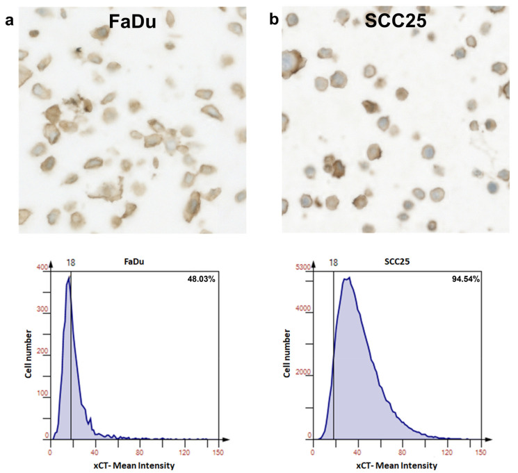
Unfavorable clinical outcomes mean that cancer researchers must attempt to develop novel therapeutic strategies to overcome therapeutic resistance in patients with HNSCC. Recently, ferroptosis was shown to be a promising pathway possessing druggable targets, such as xCT (SLC7A11). Unfortunately, little is known about the molecular mechanisms underlying the susceptibility of HNSCC cells to ferroptosis. The goal of this study was to determine whether HNSCC cells with activated Erk1/2 are vulnerable to ferroptosis induction. Our results have shown that xCT (SLC7A11) was overexpressed in malignant tissues obtained from the patients with HNSCC, whereas normal mucosa demonstrated weak expression of the protein. In order to investigate the role of Erk1/2 in the decrease in cell viability caused by erastin, xCT-overexpressing FaDu and SCC25 HNSCC cells were used. The ravoxertinib-dependent inhibition of Erk1/2 signaling led to the decrease in erastin efficacy due to the effect on ROS production and the upregulation of ROS scavengers SOD1 and SOD2, resulting in repressed lipid peroxidation. Therefore, it was concluded that the erastin-dependent activation of ferroptosis seems to be a promising approach which can be further developed as an additional strategy for the treatment of HNSCC. As ferroptosis induction via erastin is strongly dependent on the expression of Erk1/2, this MAP kinase can be considered as a predictor for cancer cells' response to erastin.

Keywords: ERK signaling; HNSCC; erastin; xCT.


Conflict of interest statement

The authors declare no conflict of interest.

Figures

Figure 1
Figure 1
xCT (SLC7A11) expression in patients with HNSCC. The prognostic value of xCT (SLC7A11) mRNA was evaluated in patients with HNSCC using the KMplot database (https://kmplot.com/analysis/), and correlation between the levels of xCT (SLC7A11) and overall survival rate is displayed on the plot diagram (a). Relative xCT (SLC7A11) mRNA levels in tissue specimens of patients with HNSCC and control healthy mucosa were determined using real-time PCR analysis (b). Relative quantities of the transcripts were calculated via pairwise differences of threshold cycles (∆CT) of the SLC7A11 gene and the loading control GAPDH housekeeping gene. *** p < 0.001.
Figure 2
Figure 2
xCT (SLC7A11) expression in normal mucosa and HNSCC samples. Immunohistochemical representation of xCT obtained via TissueFax® (TissueGnostocs™, Vienna, Austria) in normal mucosa (a) and tumor tissue (b,c); 400× magnification, bar: 50 µm (a,c); 200× magnification, bar: 100 µm (b). Differences in xCT expression in normal mucosa and HNSCC samples (d). Statistical analysis was performed with the Mann–Whitney test, *** p < 0.001.
Figure 3
Figure 3
Expression of xCT (SLC7A11) in FaDu and SCC25 HNSCC cells. Representative immunohistochemical images made using TissueFax® (TissueGnostocs™, Vienna, Austria) show the levels of the enzyme content in FaDu (a) and SCC25 (b) cells. Mean intensity was determined using the HistoQuest software.
Figure 4
Figure 4
Effects of erastin and ferrostatin-1 on viability and morphology of HNSCC cells. Cell viability was determined in FaDu and SCC25 cells at 72 h after treatment with erastin (2 µM) or combination of erastin (2 µM) and ferrostatin-1 (1 µM). DMSO was used as a vehicle control. Columns represent the mean + SEM calculated after at least three independent experiments, *** p < 0.001 (a,b) Morphological characteristics of ferroptotic cell death in HNSCC FaDu and SCC25 cells were evaluated using holotomographic microscopy (Nanolive SA 3D-Explorer-FLUO, Tolochenaz, Switzerland) as described in Materials and Methods. Cells were treated with erastin (2 µM) or a combination of erastin (2 µM) and ferrostatin-1 (1 µM), and at 72 h after treatment, the representative images were obtained; 60× magnification, bar: 20 µm.
Figure 5
Figure 5
Effects of erastin and ravoxertinib on the expression of total and phospho-Erk1/2. Levels of the total Erk1/2 (a) and phospho-Erk1 (T202 + Y204) and –Erk2 (T185 + Y187) (b) were determined in FaDu and SCC25 cells 24 h after treatment with erastin (2 µM), ravoxertinib (100 nM), or their combination. Data are presented as mean + SD after three independent experiments, * p < 0.05, ** p < 0.01.
Figure 6
Figure 6
Cell viability and lipid peroxidation in FaDu and SCC25 HNSCC cells in response to erastin and ravoxertinib exposure. FaDu (a) and SCC25 (b) cells were treated with erastin (2 µM), ravoxertinib (100 nM), or their combination, and cell viability was detected at 24, 48, and 72 h, as described in Materials and Methods. Lipid peroxidation was studied via flow cytometry analysis of the oxidized C11-BODIPY (581/591) in HNSCC cells at 24 h after treatment with erastin (2 µM), ravoxertinib (100 nM), or their combination (c). Data are given as mean + SD (a,b) or mean + SEM (c) obtained from at least three independent experiments, * p < 0.05, ** p < 0.01, *** p < 0.001.
Figure 7
Figure 7
ROS production and expression of ROS scavengers, SOD1, and SOD2 in HNSCC cells. Flow cytometry analysis of the HNSCC cells stained with DCFDA fluorogenic dye was used to measure hydroxyl, peroxyl, and other ROS activities in FaDu and SCC25 cells 24 h after treatment with erastin (2 µM), ravoxertinib (100 nM), or their combination (a). Expression levels of ROS scavengers SOD1 (b) and SOD2 (c) were evaluated using the Promega Lumit™ Immunoassay Cellular System (see Materials and Methods) 24 h after treatment with erastin (2 µM), ravoxertinib (100 nM), or their combination. Data are given as mean + SD (a) or mean + SEM (b,c) obtained from at least three independent experiments, * p < 0.05, ** p < 0.01, *** p < 0.001.
Figure 8
Figure 8
Hypothetical sequence of events leading to development of ferroptosis in HNSCC cells in response to erastin treatment. The schematic diagram illustrates that erastin inhibits xCT, resulting in downregulation of GSH, GPX4, and Nrf2 [58]. Decrease in the detoxifying molecules caused by erastin leads to enhanced ROS production, lipid peroxidation, and ferroptosis development [19,57]. EGFR pathway and its downstream Erk1/2 activation plays an important role in HNSCC pathophysiology. Erk1/2, in cooperation with Nrf2 [24], is involved in the modulation of ROS formation and expression of ROS scavengers SOD1 and SOD2. Inhibition of Erk1/2 signaling via specific Erk-inhibitor, ravoxertinib, affects the ratio between ROS levels and ROS scavengers’ expression, resulting in ferroptosis attenuation.

Similar articles

Cited by

References

    1. Sung H., Ferlay J., Siegel R.L., Laversanne M., Soerjomataram I., Jemal A., Bray F. Global cancer statistics 2020: GLOBOCAN estimates of incidence and mortality worldwide for 36 cancers in 185 countries. CA Cancer J. Clin. 2021;71:209–249. doi: 10.3322/caac.21660. - DOI - PubMed
    1. De Angelis R., Sant M., Coleman M.P., Francisci S., Baili P., Pierannunzio D., Trama A., Visser O., Brenner H., Ardanaz E. Cancer survival in Europe 1999–2007 by country and age: Results of EUROCARE-5—A population-based study. Lancet Oncol. 2014;15:23–34. doi: 10.1016/S1470-2045(13)70546-1. - DOI - PubMed
    1. Johnson D.E., Burtness B., Leemans C.R., Lui V.W.Y., Bauman J.E., Grandis J.R. Head and neck squamous cell carcinoma. Nat. Rev. Dis. Prim. 2020;6:1–22. doi: 10.1038/s41572-020-00224-3. - DOI - PMC - PubMed
    1. Raudenská M., Balvan J., Masařík M. Cell death in head and neck cancer pathogenesis and treatment. Cell Death Dis. 2021;12:192. doi: 10.1038/s41419-021-03474-5. - DOI - PMC - PubMed
    1. Dixon S.J., Lemberg K.M., Lamprecht M.R., Skouta R., Zaitsev E.M., Gleason C.E., Patel D.N., Bauer A.J., Cantley A.M., Yang W.S. Ferroptosis: An iron-dependent form of nonapoptotic cell death. Cell. 2012;149:1060–1072. doi: 10.1016/j.cell.2012.03.042. - DOI - PMC - PubMed

Publication types

MeSH terms