. 2023 Jan 12;11(1):198.
doi: 10.3390/biomedicines11010198.

Timeline of Developmental Defects Generated upon Genetic Inhibition of the Retinoic Acid Receptor Signaling Pathway

Affiliations

Timeline of Developmental Defects Generated upon Genetic Inhibition of the Retinoic Acid Receptor Signaling Pathway

Marius Teletin et al. Biomedicines. .

Abstract

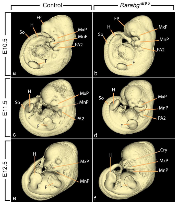
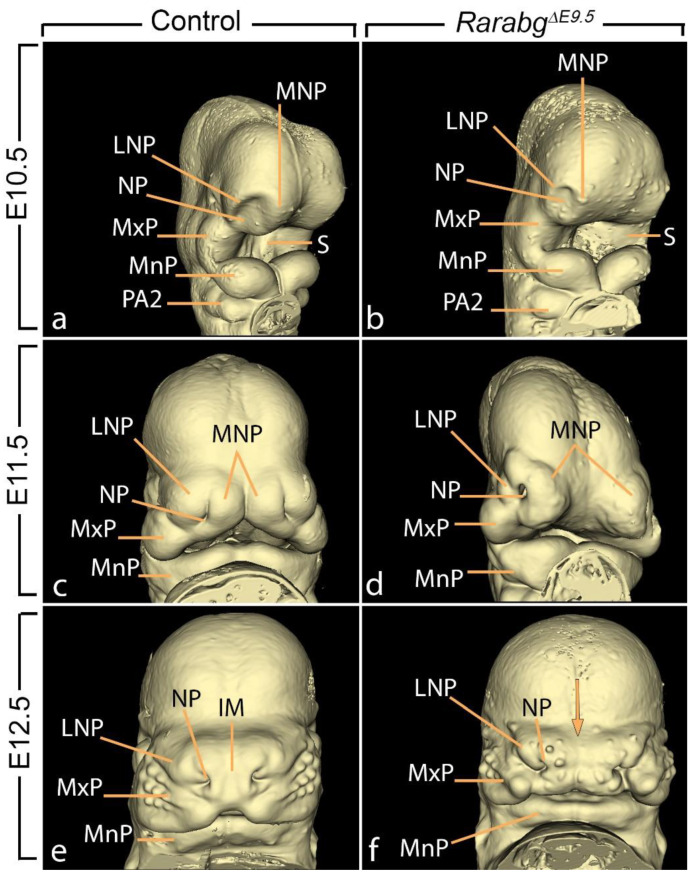
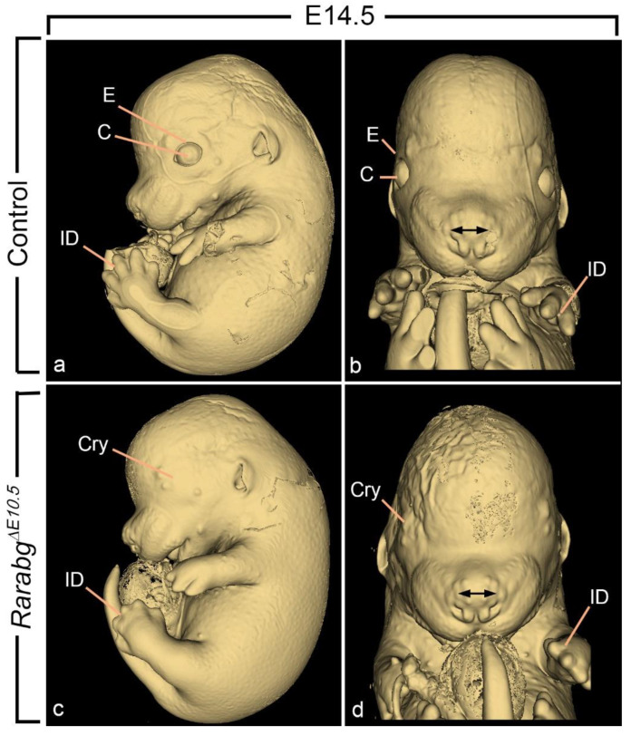
It has been established for almost 30 years that the retinoic acid receptor (RAR) signalling pathway plays essential roles in the morphogenesis of a large variety of organs and systems. Here, we used a temporally controlled genetic ablation procedure to precisely determine the time windows requiring RAR functions. Our results indicate that from E8.5 to E9.5, RAR functions are critical for the axial rotation of the embryo, the appearance of the sinus venosus, the modelling of blood vessels, and the formation of forelimb buds, lung buds, dorsal pancreatic bud, lens, and otocyst. They also reveal that E9.5 to E10.5 spans a critical developmental period during which the RARs are required for trachea formation, lung branching morphogenesis, patterning of great arteries derived from aortic arches, closure of the optic fissure, and growth of inner ear structures and of facial processes. Comparing the phenotypes of mutants lacking the 3 RARs with that of mutants deprived of all-trans retinoic acid (ATRA) synthesising enzymes establishes that cardiac looping is the earliest known morphogenetic event requiring a functional ATRA-activated RAR signalling pathway.

Keywords: HREM; axial rotation; cardiac looping; embryonic turning; eye development; heart development; inner ear development; lung development; mouse.


Conflict of interest statement

The authors declare no conflict of interest.

Figures

Figure 1
Figure 1
Correspondence between the day of TAM treatment, the time lapse for obtaining mutants with loss of RARA and RARG (colored arrows), the schedule of the phenotypic analyses, and the number of mutants (m) and control (c) embryos examined by HREM at each developmental stage. m, these mutants were incompletely excised for Rara and Rarg; see the main text for further details.
Figure 2
Figure 2
Left lateral views of control embryos (a,c,e) and RarabgΔE9.5 mutant littermates (b,d,f) at E10.5, E11.5, and E12.5, as indicated. Cry, cryptophthalmos; F, forelimb bud; FP, frontonasal process; H, hindlimb bud; MxP and MnP, maxillary and mandibular processes of the 1st pharyngeal arch, respectively; PA2, 2nd pharyngeal arch. So, somite. Same magnification in (af).
Figure 3
Figure 3
Left latero-ventral views (a,b,d,e,g) and ventral views (c,f,h) of 3D-reconstructions of the pharyngeal and foregut regions in control embryos (ac), RarabgΔE9.5 mutants (df) and RarabgΔ10.5 mutants (g,h) at E10.5, E11.5, and E12.5, as indicated. AA3, AA4, and AA6, 3rd, 4th, and 6th aortic arches; AS, aortic sac; AsA, ascending aorta, C, presumptive carina; D, duodenum; fDA, left dorsal aorta; DP, dorsal pancreatic bud; E, esophagus F, (undivided) foregut tube; G, gallbladder; rLB and fLB, lumens of the right and left lung buds, respectively; Ph, pharyngeal cavity; PP1, PP2, PP3, and PP4, 1st, 2nd, 3rd, and 4th pharyngeal pouches; PT, pulmonary trunk; PTA, persistent truncus arteriosus; rSB and fSB, right and left stem bronchi, respectively; St, stomach; T, trachea; Th, thyroid gland; VP, ventral pancreatic bud. Same magnifications in (a,d), in (b,e,g) and in (c,f,h).
Figure 4
Figure 4
Ventral views of 3D-reconstructions of the pharyngeal cavity (af) and aortic arches (df) in a control embryo (a,d) and in RarabgΔE9.5 mutants (b,c,e,f) at E10.5. AA1, AA2, AA3, AA4, and AA6, 1st, 2nd, 3rd, 4th, and 6th aortic arches; AS, aortic sac; rDA, fDA and mDA, right, left and midline dorsal aortas, respectively; rDA*, hypoplastic right dorsal aorta; PP1, PP2, PP3, and PP4, 1st, 2nd, 3rd, and 4th pharyngeal pouches. Same magnifications in (af).
Figure 5
Figure 5
Right lateral views (a,d,g), frontal views (b,e,h) and ventral views (c,f,i) of 3D-reconstructions of the head region in a control embryo (ac), in a RarabgΔE9.5 mutant (df), and in a RarabgΔ10.5 mutant (gi) at E12.5. The panel focuses: (i) on the anatomy of the optic cup and on its position relative to its neighbouring structures and (ii) on the communications between the nasal and oral cavities (the primitive choanae) in control embryos which is absent in mutant embryos, leading to choanal atresia. Ch, primitive choana; Di, cavity of the diencephalon (3rd ventricle); DR and VR, dorsal and ventral region of the retina, respectively; IC, internal carotid artery; IE, cavities of the inner ear; L, lens; N, nasal cavity; OrC, oral cavity; OD, optic disc; OF, optic fissure; OP, optic peduncle; OR, optic recess of the 3rd ventricle; P, pituitary gland; Ph, pharyngeal cavity; Tel, telencephalic vesicle. The white arrowhead points to the ventral opening in the optic cup (retinal coloboma) and the asterisks indicate the “gap” between the nasal and oral cavities resulting from the absence of the choana. Same magnifications in (a,d,g), in (b,e,h) and in (c,f,i).
Figure 6
Figure 6
Facial aspect of control embryos (a,c,e) and RarabgΔE9.5 mutant littermates (b,d,f) at E10.5, E11.5, and E12.5, as indicated. IM, intermaxillary segment; LNP and MNP, lateral and medial nasal processes, respectively; MxP and MnP, maxillary and mandibular processes of the 1st pharyngeal arch, respectively; NP, nasal pit; PA2, second pharyngeal arch; S, stomodeum (primitive mouth). The arrow (in f) points to the midfacial cleft. Same magnifications in (a,b), in (c,d) and in (e,f).
Figure 7
Figure 7
External appearance of control (a,b) and RarabgΔE10.5 mutant (b,d) littermates at E14.5. The cryptophtalmos (Cry), the failure of separation of the digits, and the shortening of the snout are characteristic of the RarabgΔE10.5 mutant phenotype (Mark et al., 2021). C, cornea; E, upper eyelid fold; ID, interdigital space. The double arrow indicates the spacing of the nostrils. Same magnifications in (a,c) and in (b,d).
Figure 8
Figure 8
Lateral views of 3D-reconstructions of the right membranous labyrinth in control embryos (a,c), in a RarabgΔE9.5 mutant (b) and in a RarabgΔ10.5 mutant (d) at E12.5 and E14.5, as indicated. The insets in (a,c) represent lateral views of normal otocysts at E9.5 and E10.5, respectively. CC, common crus; CO, cochlea; ED, endolymphatic duct; ES, endolymphatic sac; LS, lateral semicircular canal; PS, posterior semicircular canal; SA, saccule; SS, superior semicircular canal; UT, utricle. Same magnification in (ad).
Figure 9
Figure 9
Right latero-ventral views of the external and internal aspects of a control embryo (a) and of a RarabgΔE8.5 mutant littermate (b) at E9.5. AA1 and AA2, 1st and 2nd aortic arches; ACV, anterior cardinal vein; CL, cardiac loop; rDA, right dorsal aorta; F, forelimb bud; FB, forebrain; FG, foregut; FP, frontonasal process; HG, hindgut; HP, heart prominence; OA, omphalomesenteric (vitelline) artery; OV, optic vesicle; PA1 and PA2, 1st and 2nd pharyngeal arches; PP1 and PP2, 1st and 2nd pharyngeal pouches; UV, umbilical vein; SV, sinus venosus. Same magnification in (a,b).
Figure 10
Figure 10
Frontal views (a,c) and right lateral views (b,d) of 3D-reconstructions of the heart region (encircled on the insets) of a control embryo (a,b) and of a RarabgΔE8.5 mutant littermate (c,d) at E9.5. A, primitive atrium; AA1 and AA2, 1st and 2nd aortic arches, respectively; fACV and rACV, left and right anterior cardinal veins, respectively; rCCV, right common cardinal vein; fDA and rDA, left and right dorsal aortas, respectively; FP, frontonasal process; fSV and rSV, left and right horns of the sinus venosus; fUV and rUV, left and right umbilical veins, respectively; V, primitive ventricle. The asterisk indicates an aorto-atrial fistula. Same magnification in (ad).
Figure 11
Figure 11
Anatomical features characteristic of the RarabgiΔE8.5 mutant phenotype at E10.5. (a) Left lateral view of the external aspect. Note the small size of the forelimb bud (F), as opposed to the normal appearance of the hindlimb bud (H) and compare with Figure 2a. (b) Left lateral view of a 3D-reconstruction of the pharyngeal and foregut regions. Compare with Figure 3a and note: (i) the abnormal persistence of the 2nd aortic arch (AA2) and (ii) the absence of the dorsal portion of the 3rd aortic arch (AA3), of the 6th aortic arch, of the 3rd and 4th pharyngeal pouches, and of the dorsal pancreatic bud. (c,d) Ventral views of 3D-reconstructions of the brain region of a control embryo (c) and of a RarabgiΔE8.5 mutant (d) at E10.5. Note the marked hypoplasia of the mutant otocysts (O), the presence of ectopic otocysts (O*), and the absence of the lens (L) on the right side. AA2, AA3, and AA4, 2nd, 3rd, and 4th aortic arches; AS, aortic sac; fDA, left dorsal aorta; D, duodenum; Mes, cavity of the mesencephalon; OC, optic cup; Ph, pharyngeal cavity; PP1 and PP2, 1st and 2nd pharyngeal pouches; Rho, cavity of the rhombencephalon; St, stomach; VB, ventral bud of the foregut; Tel, telencephalic vesicle. Same magnifications in (c,d).
Figure 12
Figure 12
Critical periods in development requiring a functional RAR signalling pathway. The developmental processes analysed in RarabgΔE8.5, RarabgΔE9.5, RarabgΔE10.5 and RarabgΔE11.5 mutants are listed. Solid bars indicate the periods when impaired signalling through RARs causes major structural defects and stippled bars indicate the periods when the defects appear less frequent or less severe. For further details, see the main text and [4].

References

    1. Mark M., Ghyselinck N.B., Chambon P. Function of retinoic acid receptors during embryonic development. Nucl. Recept. Signal. 2009;7:e002. doi: 10.1621/nrs.07002. - DOI - PMC - PubMed
    1. Mark M., Ghyselinck N.B., Chambon P. Function of retinoid nuclear receptors: Lessons from genetic and pharmacological dissections of the retinoic acid signaling pathway during mouse embryogenesis. Annu. Rev. Pharmacol. Toxicol. 2006;46:451–480. doi: 10.1146/annurev.pharmtox.46.120604.141156. - DOI - PubMed
    1. Vernet N., Condrea D., Mayere C., Féret B., Klopfenstein M., Magnant W., Alunni V., Teletin M., Souali-Crespo S., Nef S., et al. Meiosis occurs normally in the fetal ovary of mice lacking all retinoic acid receptors. Sci. Adv. 2020;6:eaaz1139. doi: 10.1126/sciadv.aaz1139. - DOI - PMC - PubMed
    1. Mark M., Teletin M., Wendling O., Vonesch J.L., Féret B., Hérault Y., Ghyselinck N.B. Pathogenesis of anorectal malformations in retinoic acid receptor knockout mice studied by HREM. Biomedicines. 2021;9:742. doi: 10.3390/biomedicines9070742. - DOI - PMC - PubMed
    1. Ruzankina Y., Pinzon-Guzman C., Asare A., Ong T., Pontano L., Cotsarelis G., Zediak V.P., Velez M., Bhandoola A., Brown E.J. Deletion of the developmentally essential gene ATR in adult mice leads to age-related phenotypes and stem cell loss. Cell Stem Cell. 2007;1:113–126. doi: 10.1016/j.stem.2007.03.002. - DOI - PMC - PubMed

LinkOut - more resources