. 2022 Dec 20;14(1):2.
doi: 10.3390/genes14010002.

Molecular Characterization of Tropomyosin and Its Potential Involvement in Muscle Contraction in Pacific Abalone

Affiliations

Molecular Characterization of Tropomyosin and Its Potential Involvement in Muscle Contraction in Pacific Abalone

Md Abu Hanif et al. Genes (Basel). .

Abstract

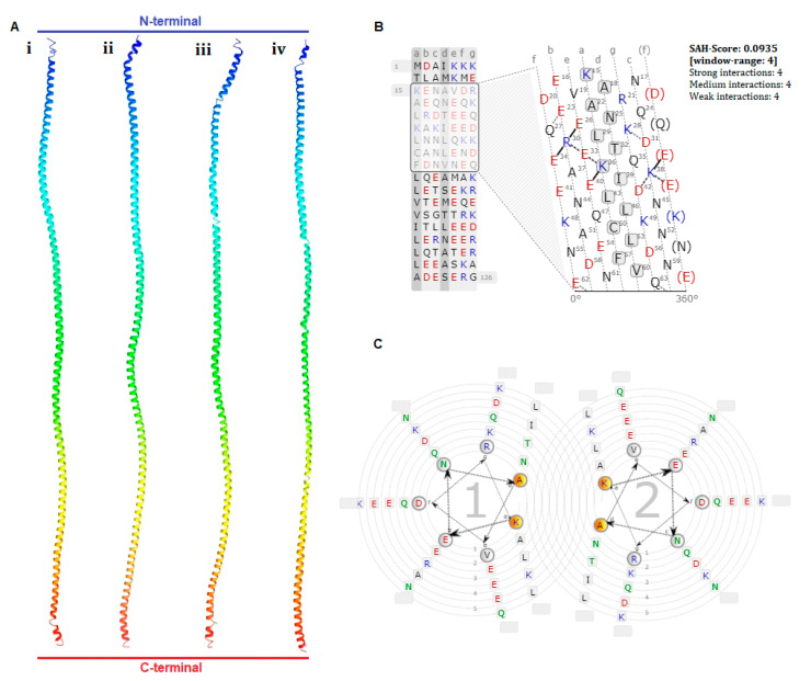
Tropomyosin (TPM) is a contractile protein responsible for muscle contraction through its actin-binding activity. The complete sequence of TPM in Haliotis discus hannai (Hdh-TPM) was 2160 bp, encoding 284 amino acids, and contained a TPM signature motif and a TPM domain. Gene ontology (GO) analysis based on the amino acid sequence predicted Hdh-TPM to have an actin-binding function in the cytoskeleton. The 3D analysis predicted the Hdh-TPM to have a coiled-coil α-helical structure. Phylogenetically, Hdh-TPM formed a cluster with other TPM/TPM1 proteins during analysis. The tissue-specific mRNA expression analysis found the higher expression of Hdh-TPM in the heart and muscles; however, during embryonic and larval development (ELD), the higher expression was found in the trochophore larvae and veliger larvae. Hdh-TPM expression was upregulated in fast-growing abalone. Increasing thermal stress over a long period decreased Hdh-TPM expression. Long-term starvation (>1 week) reduced the mRNA expression of Hdh-TPM in muscle; however, the mRNA expression of Hdh-TPM was significantly higher in the mantle, which may indicate overexpression. This study is the first comprehensive study to characterize the Hdh-TPM gene in Pacific abalone and to report the expression of Hdh-TPM in different organs, and during ELD, different growth patterns, thermal stress, seasonal changes, and starvation.

Keywords: cloning; coiled-coil structure; contractile function; expression analysis; gene ontology.


Conflict of interest statement

All authors have no conflict of interests relevant to this study to disclose.

Figures

Figure 1
Figure 1
Full-length nucleotide and deduced amino acid sequences of Hdh-TPM (GenBank accession no. OM937906.1). The numerical numbers at the left and right side of the sequence indicate the position of nucleotide and amino acid of the Hdh-TPM gene, respectively. The coding region starts with a start codon (ATG) and ends with a stop codon (TAA), shown by green bold underline letter. The tropomyosin domain is marked with blue letters. The conserved tropomyosin signature peptide is marked with a light blue box. Cyclic adenosine monophosphate (cAMP) and cyclic guanosine monophosphate (cGMP)-dependent protein kinase phosphorylation sites are denoted by a yellow box. Potential protein kinase C phosphorylation sites are marked with a green box. Predicted casein kinase II phosphorylation sites are marked with an orange box. cAMP- and cGMP-dependent protein kinase phosphorylation sites are boxed in yellow. An amidation present in the sequence is shown with a pink box. The potential glutamic-acid-rich region profile is marked with a red dotted underline. N-terminal helix-turn-helix (HTH) site is indicated by an ash box. The putative polyadenylation signal is denoted by a violet letter with a plain underline.
Figure 2
Figure 2
Schematic diagram of the motifs detected in Hdh-TPM and other TPM of invertebrates and vertebrates. Distinct motifs are denoted by different colors. The motif analysis included the TPM sequence of H. discus hannai and TPM sequences of other species: H. discus discus (BAH10148.1), H. rufescens (CAA53028.1), Crassostrea gigus (ARX70262.1), Mizuhopecten yessoensis (ACF22883.1), Scylla serrata (ABS12233.1), Pennahia argentata (BAB20881.1), Rattus norvegicus (AAA42263.1), Gallus gallus (NP_990732.1), and Homo sapiens (AAT68295.1).
Figure 3
Figure 3
The multiple sequence alignment of Hdh-TPM from deduced amino acid sequences of H. discus hannai (OM937906), H. rufescens (X75218.1), H. discus discus (AB444939.1), Crassostrea gigus (AB444943.1), Mizuhopecten yessoensis (AB004636.1), Scylla serrata (EF672351.1), Pennahia argentata (AB045645.2), Mus musculus (AAI32038.1), Gallus gallus (AAA49112.1), and Homo sapiens (AAA36771.1). Tropomyosin signature is indicated by a black outlined box.
Figure 4
Figure 4
(A) Three-dimensional structure prediction of amino acid sequence from four abalone species (i) H. discus hannai, (ii), H. asisina, (iii) H. discus discus and (iv) H. rufescens. (B) A single α helical (SAH) interaction in the protein heptad net view. (C) A coiled-coil wheel view of the Hdh-TPM protein.
Figure 5
Figure 5
Gene ontology based on the Hdh-TPM protein sequence: (A) Molecular function indicating actin-binding cytoskeletal protein. (B) Biological processes, including single-organism cellular process through cellular component organization and development. (C) Cellular component indicating intracellular organelle, especially the cytoskeleton.
Figure 6
Figure 6
Phylogenetic tree constructed using the maximum likelihood method with a bootstrap value of 1000 after clustalW alignment based on amino acid residues of different isoforms of tropomyosin. The following sequences with their protein ID were used to construct the phylogenetic tree: TPM/TPM1 of H. discus discus (BAH10148.1), H. discus hannai (AAR45321.1), H. rufescens (CAA53028.1), Crassostrea gigus (ARX70262.1), Mizuhopecten yessoensis (BAA20455.1), Mytilus galloprovincialis (VDI44733.1), Scylla serrata (ABS12233.1), and Penaeus monodon (ADV17340.1); TPM3 of Sus scrofa (NP_001001632.1), Bos taurus (NP_001011674.1), Gallus gallus (ATC20309.1), Mus musculus (AAG38596.1), Homo sapiens (ATC20307.1), and Danio rerio (NP_991239.1); TPM4 of Danio rerio (AAH53144.1), Xenopus tropicalis (CAJ82227.1), Sus scrofa (NP_999500.1), Mus musculus (AAH23701.1), Gallus gallus (XP_046789950.1), and Homo sapiens (NP_001138632.1); TPM2 of Danio rerio (XP_005155551.1), Xenopus tropicalis (NP_001025587.1), Gallus gallus (XP_046790843.1), Sus scrofa (NP_001123419.1), Homo sapiens (KAI2552574.1), and Mus musculus (NP_001264805.1).
Figure 7
Figure 7
Expression of Hdh-TPM mRNA in different tissues of Pacific abalone H. discus hannai: (A) semiquantitative expression; (B) quantitative expression. Significantly different levels (p < 0.05) are denoted by different letters.
Figure 8
Figure 8
Expression of Hdh-TPM mRNA in embryonic and larval developmental stages of Pacific abalone. Significantly different levels (p < 0.05) are denoted by different letters.
Figure 9
Figure 9
Expression of Hdh-TPM in the mantle and muscle tissues of different-growth-type Pacific abalones, H. discus hannai. Significantly different levels (p < 0.05) are denoted by different letters.
Figure 10
Figure 10
Expression of Hdh-TPM mRNA at different heat stress conditions: (A) Muscle tissue and (B) mantle tissue of Pacific abalone. Significantly different levels (p < 0.05) are denoted by different letters.
Figure 11
Figure 11
Expression of Hdh-TPM according to the season in mantle and muscle tissues of Pacific abalone H. discus hannai. Significantly different levels (p < 0.05) are denoted by different letters.
Figure 12
Figure 12
Expression of Hdh-TPM in the mantle and muscle of starved Pacific abalone H. discus hannai. Significantly different levels (p < 0.05) are denoted by different letters.

Similar articles

References

    1. Kalyva A., Schmidtmann A., Geeves M.A. In vitro formation and characterization of the skeletal muscle α·β Tropomyosin heterodimers. Biochemistry. 2012;51:6388–6399. doi: 10.1021/bi300340r. - DOI - PMC - PubMed
    1. Gunning P.W., Ghoshdastider U., Whitaker S., Popp D., Robinson R.C. The evolution of compositionally and functionally distinct actin filaments. J. Cell Sci. 2015;128:2009–2019. doi: 10.1242/jcs.165563. - DOI - PubMed
    1. Behrmann E., Muller M., Penczek P.A., Mannherz H.G., Manstein D.J., Raunser S. Structure of the rigor actin-Tropomyosin-myosin complex. Cell. 2012;150:327–338. doi: 10.1016/j.cell.2012.05.037. - DOI - PMC - PubMed
    1. Barua B., Winkelmann D.A., White H.D., Hitchcock-DeGregori S.E. Regulation of actin-myosin interaction by conserved periodic sites of Tropomyosin. Proc. Natl. Acad. Sci. USA. 2012;109:18425–18430. doi: 10.1073/pnas.1212754109. - DOI - PMC - PubMed
    1. Loong C.K., Badr M.A., Chase P.B. Tropomyosin flexural rigidity and single Ca2+ regulatory unit dynamics: Implications for cooperative regulation of cardiac muscle contraction and cardiomyocyte hypertrophy. Front. Physiol. 2012;4:80. doi: 10.3389/fphys.2012.00080. - DOI - PMC - PubMed

Publication types

LinkOut - more resources