. 2022 Dec 26;14(1):74.
doi: 10.3390/genes14010074.

Genetic Diagnosis for 64 Patients with Inherited Retinal Disease

Affiliations

Genetic Diagnosis for 64 Patients with Inherited Retinal Disease

Jacob Lynn et al. Genes (Basel). .

Abstract

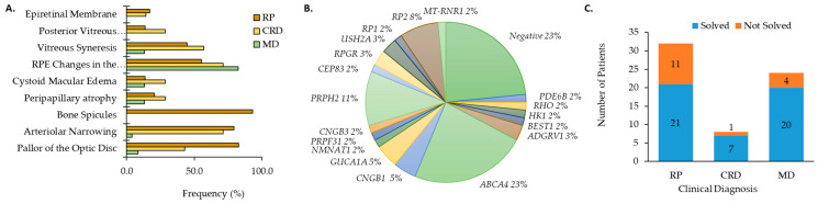
The overlapping genetic and clinical spectrum in inherited retinal degeneration (IRD) creates challenges for accurate diagnoses. The goal of this work was to determine the genetic diagnosis and clinical features for patients diagnosed with an IRD. After signing informed consent, peripheral blood or saliva was collected from 64 patients diagnosed with an IRD. Genetic testing was performed on each patient in a Clinical Laboratory Improvement Amendments of 1988 (CLIA) certified laboratory. Mutations were verified with Sanger sequencing and segregation analysis when possible. Visual acuity was measured with a traditional Snellen chart and converted to a logarithm of minimal angle of resolution (logMAR). Fundus images of dilated eyes were acquired with the Optos® camera (Dunfermline, UK). Horizontal line scans were obtained with spectral-domain optical coherence tomography (SDOCT; Spectralis, Heidelberg, Germany). Genetic testing combined with segregation analysis resolved molecular and clinical diagnoses for 75% of patients. Ten novel mutations were found and unique genotype phenotype associations were made for the genes RP2 and CEP83. Collective knowledge is thereby expanded of the genetic basis and phenotypic correlation in IRD.

Keywords: clinical; genetic testing; inherited retinal disease.


Conflict of interest statement

The authors declare no conflict of interest.

Figures

Figure 1
Figure 1
Clinical characteristics for patients with (A) cone rod dystrophy (CRD) and retinitis pigmentosa (RP) and macular dystrophy (MD). (B) Distribution of genes causative for ocular disease by number of patients (%). (C) The number of genetically solved cases subset by clinical diagnosis.
Figure 2
Figure 2
Phenotype of two families with X-linked RP (XLRP) due to mutations in RP2. (A) Pedigree analysis for patient GP006 showed three generations of RP (black squares). (B) Fundus images for GP006 showed optic disc pallor OU, arteriolar narrowing, and bone spicules in the peripheral retina. (C) Fundus autofluorescence (FAF) revealed hypo-AF in the nasal periphery and fovea. (D) SDOCT showed ellipsoid zone (EZ) changes and retinal pigment epithelium (RPE) atrophy OU. GP006a (EG) had typical RP features (E) with hypo-AF regions in the nasal retina and throughout the posterior pole (F) and thinning of the outer retina (G). GP006b (HJ) had pallor of the optic disc, chorioretinal atrophy of the far periphery and pigment migration in the inferior retina (H). Radial lines and inferior hypo-AF were noted on FAF (I). SDOCT (J) showed outer retinal atrophy in the temporal macula OU for GP006b. (K) Pedigree analysis for patient GP063 indicated maternally inherited RP with affected family members from three generations. GP063 (LN) had typical RP features, an atrophic macula, and a loss of EZ band on SDOCT. GP063a (OQ) had typical RP, multifocal areas of hypo-AF, a loss of EZ temporally OD (upper panel), and complete loss OS (lower panel).
Figure 3
Figure 3
Phenotype associated with CEP83 mutations. (A) Fundus images for GP037 were showing chorioretinal atrophy, RPE changes in the macula and typical RP features. (B) Fundus autofluorescence imaging showed an atrophic midperiphery and a bullseye pattern of autofluorescence. (C) SDOCT imaging was notable for outer retinal atrophy with foveal preservation of the ellipsoid zone (EZ) line.

Similar articles

Cited by

References

    1. Chan T., Friedman D.S., Bradley C., Massof R. Estimates of incidence and prevalence of visual impairment, low vision, and blindness in the united states. JAMA Ophthalmol. 2018;136:12–19. doi: 10.1001/jamaophthalmol.2017.4655. - DOI - PMC - PubMed
    1. Bessant D.A., Ali R.R., Bhattacharya S.S. Molecular genetics and prospects for therapy of the inherited retinal dystrophies. Curr. Opin. Genet. Dev. 2001;11:307–316. doi: 10.1016/S0959-437X(00)00195-7. - DOI - PubMed
    1. Sohocki M.M., Daiger S.P., Bowne S.J., Rodriquez J.A., Northrup H., Heckenlively J.R., Birch D., Mintz-Hittner H., Ruiz R.S., Lewis R.A., et al. Prevalence of mutations causing retinitis pigmentosa and other inherited retinopathies. Hum. Mutat. 2001;17:42–51. doi: 10.1002/1098-1004(2001)17:1<42::AID-HUMU5>3.0.CO;2-K. - DOI - PMC - PubMed
    1. Fahim A.T., Daiger S.P., Weleber R.G. Nonsyndromic retinitis pigmentosa overview. In: Adam M.P., Ardinger H.H., Pagon R.A., Wallace S.E., Bean L.J.H., Stephens K., Amemiya A., editors. Genereviews((r)) University of Washington; Seattle, WA, USA: 1993. - PubMed
    1. Daiger S.P., Sullivan L.S., Bowne S.J. Genes and mutations causing retinitis pigmentosa. Clin. Genet. 2013;84:132–141. doi: 10.1111/cge.12203. - DOI - PMC - PubMed

Publication types

LinkOut - more resources