. 2023 Jan 4;13(2):179.
doi: 10.3390/diagnostics13020179.

High-Frequency (30 MHz-6 GHz) Breast Tissue Characterization Stabilized by Suction Force for Intraoperative Tumor Margin Assessment

Affiliations

High-Frequency (30 MHz-6 GHz) Breast Tissue Characterization Stabilized by Suction Force for Intraoperative Tumor Margin Assessment

Hadi Mokhtari Dowlatabad et al. Diagnostics (Basel). .

Abstract

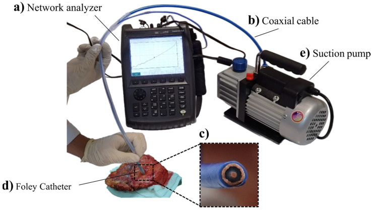
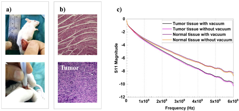
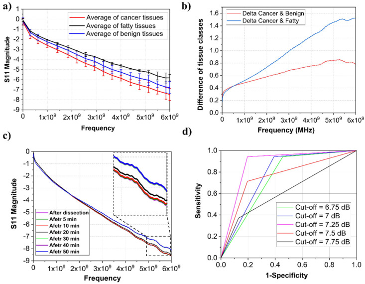
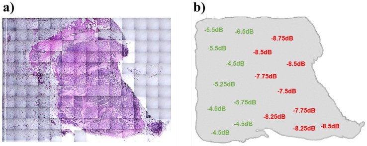
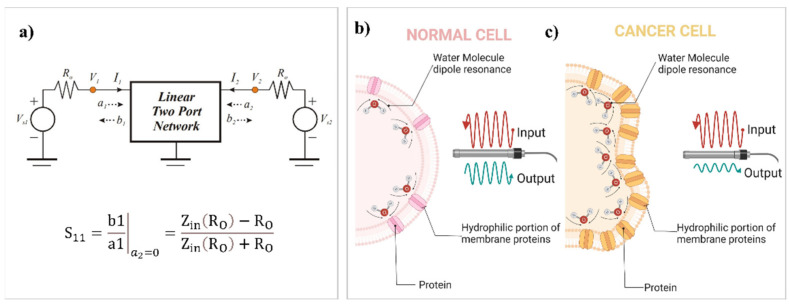
A gigahertz (GHz) range antenna formed by a coaxial probe has been applied for sensing cancerous breast lesions in the scanning platform with the assistance of a suction tube. The sensor structure was a planar central layer and a metallic sheath of size of 3 cm2 connected to a network analyzer (keySight FieldFox N9918A) with operational bandwidth up to 26.5 GHz. Cancer tumor cells have significantly higher water content (as a dipolar molecule) than normal breast cells, changing their polarization responses and dielectric losses to incoming GHz-based stimulation. Principal component analysis named S11, related to the dispersion ratio of the input signal, is used as a parameter to identify malignant tumor cells in a mouse model (in vivo) and tumor specimens of breast cancer patients (in vitro) (both central and marginal parts). The results showed that S11 values in the frequency range from 5 to 6 GHz were significantly higher in cancer-involved breast lesions. Histopathological analysis was the gold standard for achieving the S11 calibration to distinguish normal from cancerous lesions. Our calibration on tumor specimens presented 82% positive predictive value (PPV), 100% negative predictive value (NPV), and 86% accuracy. Our goal is to apply this system as an in vivo non-invasive tumor margin scanner after further investigations in the future.

Keywords: GHz spectroscopy; breast cancer; dipolar polarization; scattering; tumor margin.


Conflict of interest statement

All the authors declare that they have no known competing financial interests or personal relationships that could have influenced the work reported in this paper.

Figures

Figure 1
Figure 1
GHz measurement system. (a) The used network analyzer (KeySight FieldFox N9918A). (b) The open-ended coaxial cable (Semi-Flexible Cable 670-086 SXE). (c) Magnified view of the coaxial cable. (d) A Foley catheter modified for applying suction thoroughly. (e) Suction pump.
Figure 2
Figure 2
GHz measurement on a mouse model. (a) Tumor tissue and applying the GHz probe on it. (b) Normal mouse tissue with H&E assay representing the fibrotic connective tissue and cancerous mouse tissue with H&E assay representing a hypercellular region and high nucleus-to-cytoplasm size ratio. (c) Frequency-dependent behaviour of S11 parameter for normal and cancerous tissues with or without applying suction force.
Figure 3
Figure 3
(a) mean values of S11 magnitude in three categories of fatty, benign, and malignant breast tissues. (b) Differences of each category versus tumor spectrum. (c) Variation of measured S11 by the time after dissection with magnification. (d) The ROC curve for different cut-offs.
Figure 4
Figure 4
GHz spectroscopy on human tumor margins. (a) H&E assay of a measured margin sample, 2.7 × 2.7 cm. (b) S11 magnitude measurements on the selected margin in 18 points. Pathologically negative and positive points are represented in green and red, respectively. The obtained pattern is completely matched with the H&E assay.
Figure 5
Figure 5
(a) Schematic view of an electrical network with two ports in a high-frequency region to describe scattering parameter, S11. (b) Schematic view of protein content and water distribution of a normal cell, which results in an increased S11 parameter in normal tissue due to less resonant membrane-bound waters associated with decreased expression of membrane proteins. (c) Schematic view of protein content and water distribution of a cancer cell, which results in a decreased S11 parameter in cancerous tissue due to more resonant membrane-bound waters associated with increased expression of membrane proteins.

References

    1. Maloney B.W., McClatchy D.M., Pogue B.W., Paulsen K.D., Wells W.A., Barth R.J. Review of Methods for Intraoperative Margin Detection for Breast Conserving Surgery. J. Biomed. Opt. 2018;23:100901. doi: 10.1117/1.JBO.23.10.100901. - DOI - PMC - PubMed
    1. Tummers Q.R.J.G., Verbeek F.P.R., Schaafsma B.E., Boonstra M.C., Van Der Vorst J.R., Liefers G.J., Van De Velde C.J.H., Frangioni J.V., Vahrmeijer A.L. Real-Time Intraoperative Detection of Breast Cancer Using near-Infrared Fluorescence Imaging and Methylene Blue. Eur. J. Surg. Oncol. 2014;40:850–858. doi: 10.1016/j.ejso.2014.02.225. - DOI - PMC - PubMed
    1. Jaafar H. Intra-Operative Frozen Section Consultation: Concepts, Applications and Limitations. Malays. J. Med. Sci. 2006;13:4–12. - PMC - PubMed
    1. Taxy J.B. Frozen Section and the Surgical Pathologist a Point of View. Arch. Pathol. Lab. Med. 2009;133:1135–1138. doi: 10.5858/133.7.1135. - DOI - PubMed
    1. Du Z., Wan H., Chen Y., Pu Y., Wang X. Bioimpedance Spectroscopy Can Precisely Discriminate Human Breast Carcinoma from Benign Tumors. Medicine. 2017;96:e5970. doi: 10.1097/MD.0000000000005970. - DOI - PMC - PubMed

LinkOut - more resources