

Giant Gallbladder Tumor, Unusual Cancer-Case Report and Short Review of Literature
- PMID: 36673003
- PMCID: PMC9857600
- DOI: 10.3390/diagnostics13020194
Giant Gallbladder Tumor, Unusual Cancer-Case Report and Short Review of Literature
Abstract
Background: Giant gallbladder is an uncommon condition that can result from a benign pathology and rarely presents with malignancy. Intracholecystic papillary-tubular neoplasm (ICPN) is a relatively new entity first described by V. Adsay in 2012 and included in the World Health Classification of Digestive System Tumours in 2019. Intracholecystic papillary-tubular neoplasm is a preinvasive lesion with an incidence of around 1% that may present as four histologic subtypes-biliary, gastric, intestinal, or oncocytic-of which the biliary subtype has the highest risk of associated invasive cancer. Although invasive carcinoma is present in about 50% of cases of ICPN, these patients have a significantly better prognosis than those with usual gallbladder cancer, suggesting that the entities may have distinct biological signatures.
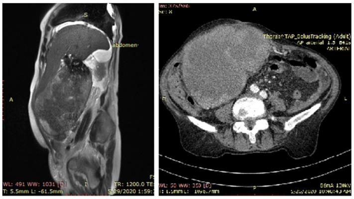
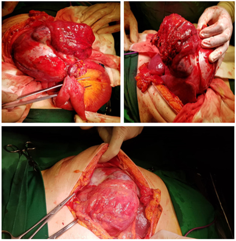
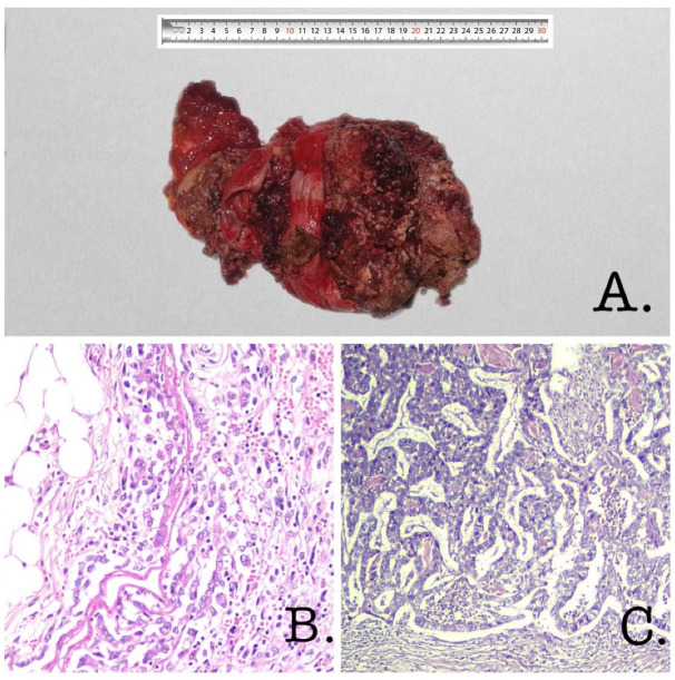
Case report: A 77-year-old female presented to the hospital with progressive swelling in the right hemiabdomen, a loss of appetite, and weight loss. MRI highlighted a giant abdominal tumor located in the right hypochondrium and right abdominal flank with liver invasion (segment V). Preoperatively, a gallbladder 25 × 17 cm in size was noted, and the patient underwent radical cholecystectomy. It was surprising to find such a giant malignant gallbladder tumor, diagnosed as invasive poorly cohesive carcinoma associated with ICPN.
Discussion: A megacholecyst is a rare discovery. Although most often found in benign pathologies, giant gallbladder cancer can be considered. The neoplastic features and the loco-regional extension of the tumor must be evaluated by imaging scans. Few cases of giant benign gallbladder have been reported in the literature; however, this appeared to be the largest resectable gallbladder carcinoma reported to date according to the literature.
Conclusion: The stage of gallbladder neoplasia is not correlated with the size of the gallbladder. Regardless of tumor size, the prognosis seems to be directly related to the stage, morphology, and resectability.
Keywords: cholecystectomy; gallbladder cancer; giant gallbladder; intracholecystic papillary–tubular neoplasm.
Conflict of interest statement
The authors declare no conflict of interest. All authors have completed the ICMJE uniform disclosure form.
Figures
Similar articles
-
Intracholecystic papillary-tubular neoplasms (ICPN) of the gallbladder (neoplastic polyps, adenomas, and papillary neoplasms that are ≥1.0 cm): clinicopathologic and immunohistochemical analysis of 123 cases.Am J Surg Pathol. 2012 Sep;36(9):1279-301. doi: 10.1097/PAS.0b013e318262787c. Am J Surg Pathol. 2012. PMID: 22895264
-
Intracholecystic papillary neoplasm associated with invasive carcinoma of the remnant gallbladder after subtotal cholecystectomy: a case report.Surg Case Rep. 2022 Feb 21;8(1):31. doi: 10.1186/s40792-022-01388-8. Surg Case Rep. 2022. PMID: 35187597 Free PMC article.
-
Three cases of intracystic papillary neoplasm of gallbladder.Abdom Radiol (NY). 2018 Jul;43(7):1535-1539. doi: 10.1007/s00261-018-1595-z. Abdom Radiol (NY). 2018. PMID: 29623349
-
The Clinical and Pathologic Features of Intracholecystic Papillary-Tubular Neoplasms of the Gallbladder.Surg Pathol Clin. 2022 Sep;15(3):565-577. doi: 10.1016/j.path.2022.05.011. Surg Pathol Clin. 2022. PMID: 36049837 Review.
-
Intracholecystic Papillary-tubular Neoplasm (ICPN) of the Gallbladder: A Case Report Focusing on an Unexpected Pathological Finding.Recent Pat Anticancer Drug Discov. 2025;20(1):131-135. doi: 10.2174/0115748928256837231012151452. Recent Pat Anticancer Drug Discov. 2025. PMID: 37885107 Review.
References
Publication types
LinkOut - more resources
Full Text Sources
