Review
. 2023 Jan 7;13(2):225.
doi: 10.3390/diagnostics13020225.

Adult Acquired Flatfoot Deformity: A Narrative Review about Imaging Findings

Affiliations
Review

Adult Acquired Flatfoot Deformity: A Narrative Review about Imaging Findings

Chiara Polichetti et al. Diagnostics (Basel). .

Abstract

Adult acquired flatfoot deformity (AAFD) is a disorder caused by repetitive overloading, which leads to progressive posterior tibialis tendon (PTT) insufficiency. It mainly affects middle-aged women and occurs with foot pain, malalignment, and loss of function. After clinical examination, imaging plays a key role in the diagnosis and management of this pathology. Imaging allows confirmation of the diagnosis, monitoring of the disorder, outcome assessment and complication identification. Weight-bearing radiography of the foot and ankle are gold standard for the diagnosis of AAFD. Magnetic Resonance Imaging (MRI) is not routinely needed for the diagnosis; however, it can be used to evaluate the spring ligament and the degree of PTT damage which can help to guide surgical plans and management in patients with severe deformity. Ultrasonography (US) can be considered another helpful tool to evaluate the condition of the PTT and other soft-tissue structures. Computed Tomography (CT) provides enhanced, detailed visualization of the hindfoot, and it is useful both in the evaluation of bone abnormalities and in the accurate evaluation of measurements useful for diagnosis and post-surgical follow-up. Other state-of-the-art imaging examinations, like multiplanar weight-bearing imaging, are emerging as techniques for diagnosis and preoperative planning but are not yet standardized and their scope of application is not yet well defined. The aim of this review, performed through Pubmed and Web of Science databases, was to analyze the literature relating to the role of imaging in the diagnosis and treatment of AAFD.

Keywords: adult acquired flatfoot deformity; foot and ankle; imaging; posterior tibial tendon; progressive collapsing foot deformity; weight bearing CT.


Conflict of interest statement

The authors declare no conflict of interest.

Figures

Figure 1
Figure 1
Taler-first metatarsal angle in the anteroposterior view (between the long axis of the talus and the long axis of the first metatarsal). (a) Normal foot, 3°; (b) pathological flatfoot, 30°.
Figure 2
Figure 2
Talonavicular coverage angle (between the line that joins the medial and lateral articular margins of the talus, and the line between that joins the medial and lateral articular margins of the navicular). (a) Normal foot, 2°; (b) pathological flatfoot, 35°.
Figure 3
Figure 3
Talonavicular uncoverage percentage (percentage of the talus that is not in contact with the navicular medially). (a) Normal foot, <30%; (b) pathological flatfoot, >30%.
Figure 4
Figure 4
Talar incongruency angle (formed by the intersection between a line from the most lateral point of the articular surfaces of the talus and the navicular, and a line from the lateral aspect of the talar neck (in its most narrow segment) to the lateral point of the talar articular surface). (a) Normal foot, 6°; (b) pathological flatfoot, >30°.
Figure 5
Figure 5
Talar-first metatarsal angle in the lateral view (Meary’s angle, the angle between the long axis of the talus and the long axis of the first metatarsal). (a) Normal foot, 4°; (b) pathological flatfoot, 30°.
Figure 6
Figure 6
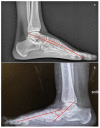
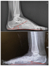
Calcaneal pitch (formed by the line parallel to the ground and the line along the inferior inclination axis of the calcaneus) in the lateral X-ray view. (a) Normal foot, 30°; (b) pathological flatfoot, 10°.
Figure 7
Figure 7
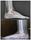
Talocalcaneal angle (formed by the long axis of the rearfoot and the midtalar line) in lateral X-ray view. (a) Normal foot, 45°; (b) pathological flatfoot, 52°.
Figure 8
Figure 8
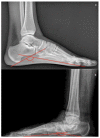
Calcaneal-fifth metatarsal angle (defined as the angle formed between the tangent to the inferior aspect of the calcaneus and a line drawn along the inferior aspect of the base and head of the fifth metatarsal) in the lateral X-ray view. (a) Normal foot, 145°; (b) pathological flatfoot, 175°.
Figure 9
Figure 9
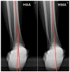
Saltzman view. (a) Hindfoot Alignment Angle (HAA) (formed by the intersection of the longitudinal axis of the tibial shaft and the axis of the calcaneal tuberosity). (b) Hindfoot Moment Arm (measured by the shortest distance between the midtibial axis and the most inferior portion of the calcaneus gus, represented by the red arrow).
Figure 10
Figure 10
Magnetic Resonance Imaging. (a) Coronal T1: soft tissue involvement of the posterior tibial tendon, (b) Axial T2: superomedial fibers of the spring ligament (white arrow).
Figure 11
Figure 11
Ultrasonography of spring ligament (marked by white arrows).
Figure 12
Figure 12
Computed Tomography (CT) imaging: (a) Talar–first metatarsal angle (Meary’s angle), (b) Medial Cuneiform-First metatarsal angle, (c) Calcaneal inclination angle, (d) Cuboid to floor distance, (e) Forefoot arch angle.

References

    1. Auch E., Barbachan Mansur N.S., Alexandre Alves T., Cychosz C., Lintz F., Godoy-Santos A.L., Baumfeld D.S., de Cesar Netto C. Distal Tibiofibular Syndesmotic Widening in Progressive Collapsing Foot Deformity. Foot Ankle Int. 2021;42:768–775. doi: 10.1177/1071100720982907. - DOI - PubMed
    1. Myerson M.S., Thordarson D.B., Johnson J.E., Hintermann B., Sangeorzan B.J., Deland J.T., Schon L.C., Ellis S.J., de Cesar Netto C. Classification and Nomenclature: Progressive Collapsing Foot Deformity. Foot Ankle Int. 2020;41:1271–1276. doi: 10.1177/1071100720950722. - DOI - PubMed
    1. de Cesar Netto C., Myerson M.S., Day J., Ellis S.J., Hintermann B., Johnson J.E., Sangeorzan B.J., Schon L.C., Thordarson D.B., Deland J.T. Consensus for the Use of Weightbearing CT in the Assessment of Progressive Collapsing Foot Deformity. Foot Ankle Int. 2020;41:1277–1282. doi: 10.1177/1071100720950734. - DOI - PubMed
    1. Bernasconi A., Ray R. Role of Minimally Invasive Surgery in Adult Flatfoot Deformity. Foot Ankle Clin. 2020;25:479–491. doi: 10.1016/j.fcl.2020.05.007. - DOI - PubMed
    1. Lee M.S., Vanore J.V., Thomas J.L., Catanzariti A.R., Kogler G., Kravitz S.R., Miller S.J., Gassen S.C. Diagnosis and Treatment of Adult Flatfoot. J. Foot Ankle Surg. 2005;44:78–113. doi: 10.1053/j.jfas.2004.12.001. - DOI - PubMed

LinkOut - more resources