. 2023 Jan 4;24(2):968.
doi: 10.3390/ijms24020968.

Identification of the Actin-Binding Region and Binding to Host Plant Apple Actin of Immunodominant Transmembrane Protein of ' Candidatus Phytoplasma mali'

Affiliations

Identification of the Actin-Binding Region and Binding to Host Plant Apple Actin of Immunodominant Transmembrane Protein of ' Candidatus Phytoplasma mali'

Kajohn Boonrod et al. Int J Mol Sci. .

Abstract

'Candidatus Phytoplasma mali' ('Ca. P. mali') has only one major membrane protein, the immunodominant membrane protein (Imp), which is regarded as being close to the ancestor of all phytoplasma immunodominant membrane proteins. Imp binds to actin and possibly facilitates its movement in the plant or insect host cells. However, protein sequences of Imp are quite diverse among phytoplasma species, thus resulting in difficulties in identifying conserved domains across species. In this work, we compare Imp protein sequences of 'Ca. P. mali' strain PM19 (Imp-PM19) with Imp of different strains of 'Ca. P. mali' and identify its actin-binding domain. Moreover, we show that Imp binds to the actin of apple (Malus x domestica), which is the host plant of 'Ca. P. mali'. Using molecular and scanning force spectroscopy analysis, we find that the actin-binding domain of Imp-PM19 contains a highly positively charged amino acid cluster. Our result could allow investigating a possible correlation between Imp variants and the infectivity of the corresponding 'Ca. P. mali' isolates.

Keywords: actin; immunodominant membrane protein; phytoplasma; single-molecule force spectroscopy.


Conflict of interest statement

The authors declare no conflict of interest.

Figures

Figure 1
Figure 1
Amino acid sequence alignment of Imp-PM19 of different ‘Ca. P. mali’strains. (a) Amino acid sequences of Imp obtained from Gene Bank were aligned using DNA star program. Color letter indicate conserved amino acids. The grey shaded area (circled) in consensus math indicates the conserved regions. (b) Protein sequence of Imp-PM19 and (c) mutants. Green, Red and Blue labeled are D1-D3 conserved regions and their pI which were mutated (underlined letters) to generate ΔD1-∆D3-Imp-PM19-mutants (italic and underlined letters), respectively.
Figure 2
Figure 2
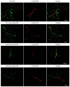
Co-expression of Imp-PM19, its mutants and mTalin in planta. Imp-PM19 and its mutants were fused with GFP and co-transiently expressed with mTalin fused to RFP to mark the plant actin. The localization of expressed proteins were analyzed by visualizing the infiltrated leaves mesophyll under confocal microscopy using GFP and RFP filters. The F-actin can be clearly seen as a long stretch filament (Red colour) in the middle panel. Co-localization of Imp-GFP and mTalin-Red is indicated by yellow colouring in the merger. A magnification of Figure 2 can be seen in Supplement Figure S1.
Figure 3
Figure 3
Coomassie blue straining SDS-PAGE of purified recombinant Imp-PM19 and ∆D3-Imp-PM19 mutant. His-tagged Imp-PM19 and ∆D3-Imp-PM19-mutant were expressed recombinantly in E. coli. The proteins were purified using Ni-NTA fast flow column. M is a protein marker. Lane 1 and 2 are purified Imp-PM19-His and ∆D3-Imp-PM19-His, respectively.
Figure 4
Figure 4
Co-sedimentation assay with recombinant actinapple, Imp-PM19 and ∆D3-Imp-PM19-mutant. (a) Coomassie-stained SDS-PAGE of recombinant apple actin. M is a protein marker. 1 is recombinant apple actin. (b) Western blot of recombinant actinapple. Recombinant actinapple (lane 1) and actinrabbit (lane 2, a positive control) were polymerized and subjected to SDS-PAGE. After blotting, the proteins were detected using anti-actin and anti-mouse-POD, respectively. (c) Labelling F-actins with Acti-stain™ fluorescent phalloidin 555 (Cytoskeleton, Inc). The labelled F-actins were visualized under a confocal microscope with a bandpass 575–615 nm filter. (d) Co-sedimentation assay. Imp-PM19 and ∆D3-Imp-PM19 mutant were incubated with F-actinapple and F-actinrabbit. After height speed centrifugation, the protein pellets were subjected to SDS-PAGE, and a western blot was performed using anti-His and anti-mouse-POD, respectively. Lane 1 and 2 are co-sedimented Imp-PM19 (1) and ∆D3-Imp-PM19-mutant (2) with F-actinrabbit. Lane 3 and 4 are co-sedimented Imp-PM19 (3) and ∆D3-Imp-PM19-mutant (4) with recombinant F-actinapple. Lane 5 and 6 are BSA (negative controls) co-sedimented with Imp-PM19 (5) and ∆D3-Imp-PM19-mutant (6). (e) An alignment of rabbit muscle actin (gene ID: 10152413) and apple actin (gene ID: XP_008355144.1). The alignment result shows 88% identity of the two actins.
Figure 5
Figure 5
Single molecule force distance curves of an Imp-PM19/∆D3-Imp-PM19 mutant functionalized SFM-tip on the F-actinrabbit surface measured by SFM. The interactions of F-actinrabbit with the Imp-PM19 (a) and the ∆D3-Imp-PM19 mutant (b) are represented.
Figure 6
Figure 6
Comparing the obtained interaction parameters with Imp-PM19 (blue bar) and the ∆D3-Imp-PM19 mutant (red bar) on F-actinrabbit filament. The work of adhesion (a) and the most frequently onserved adhesion (b) of Imp-PM19 and ∆D3-Imp-PM19 with F-actinrabbit filament ligands are measured by SMFS. The results are graphically represented. Retraction speed 2 µm/s, number of curves = 1000, set point of 800 pN.
Figure 7
Figure 7
Prediction of Imp protein folding using Phyre2 computing program. The protein sequence of Imp-PM19 was entered to Phyre2 computing program. The result shows that Imp-PM19 folds as alpha-alpha-superhelix form with confidence 17.72%. The colour scale indicates confidence score.

References

    1. Murray R.G., Stackebrandt E. Taxonomic note: Imp-PM19lementation of the provisional status Candidatus for incompletely described prokaryotes. Int. J. Syst. Bacteriol. 1995;45:186–187. doi: 10.1099/00207713-45-1-186. - DOI - PubMed
    1. IRPCM ‘Candidatus Phytoplasma’, a taxon for the wall-less, non-helical prokaryotes that colonise plant phloem and insects. J. Syst. Evol. Microbiol. 2004;54:1243–1255. doi: 10.1099/ijs.0.02854-0. - DOI - PubMed
    1. Ding L., Lu W., Yang Y., Zhong Q., Zhou T., Wang G., Song C., Ma W., Chen W., Wu Y. Immunodominant membrane protein (Imp) promotes the transmission of wheat blue dwarf (WBD) phytoplasma by directly interacting with α-tubulin in leafhoppers. Eur. J. Plant Pathol. 2022;162:357–367. doi: 10.1007/s10658-021-02408-3. - DOI
    1. Strohmayer A., Schwarz T., Braun M., Krczal G., Boonrod K. The Effect of the Anticipated Nuclear Localization Sequence of ‘Candidatus Phytoplasma mali’ SAP11-like Protein on Localization of the Protein and Destabilization of TCP Transcription Factor. Microorganisms. 2021;9:1756. doi: 10.3390/microorganisms9081756. - DOI - PMC - PubMed
    1. Clark M.F., Morton A., Buss S.L. Preparation of mycoplasma immunogens from plants and a comparison of polyclonal and monoclonal antibodies made against primula yellows MLO-associated antigens. Ann. Appl. Biol. 1989;114:111–124. doi: 10.1111/j.1744-7348.1989.tb06791.x. - DOI

LinkOut - more resources