. 2023 Jan 4;24(2):981.
doi: 10.3390/ijms24020981.

Recombinant Human Clusterin Seals Damage to the Ocular Surface Barrier in a Mouse Model of Ophthalmic Preservative-Induced Epitheliopathy

Affiliations

Recombinant Human Clusterin Seals Damage to the Ocular Surface Barrier in a Mouse Model of Ophthalmic Preservative-Induced Epitheliopathy

Shravan K Chintala et al. Int J Mol Sci. .

Abstract

There is a significant unmet need for therapeutics to treat ocular surface barrier damage, also called epitheliopathy, due to dry eye and related diseases. We recently reported that the natural tear glycoprotein CLU (clusterin), a molecular chaperone and matrix metalloproteinase inhibitor, seals and heals epitheliopathy in mice subjected to desiccating stress in a model of aqueous-deficient/evaporative dry eye. Here we investigated CLU sealing using a second model with features of ophthalmic preservative-induced dry eye. The ocular surface was stressed by topical application of the ophthalmic preservative benzalkonium chloride (BAC). Then eyes were treated with CLU and sealing was evaluated immediately by quantification of clinical dye uptake. A commercial recombinant form of human CLU (rhCLU), as well as an rhCLU form produced in our laboratory, designed to be compatible with U.S. Food and Drug Administration guidelines on current Good Manufacturing Practices (cGMP), were as effective as natural plasma-derived human CLU (pCLU) in sealing the damaged ocular surface barrier. In contrast, two other proteins found in tears: TIMP1 and LCN1 (tear lipocalin), exhibited no sealing activity. The efficacy and selectivity of rhCLU for sealing of the damaged ocular surface epithelial barrier suggests that it could be of therapeutic value in treating BAC-induced epitheliopathy and related diseases.

Keywords: clusterin; dry eye; epitheliopathy; matrix metalloproteinase inhibitor; molecular chaperone; ocular surface.


Conflict of interest statement

M.E.F. and S.J. are named as co-inventors on US patent number 9241974 entitled “Clusterin Pharmaceuticals and Treatment Methods Using the Same” granted to the University of Southern California. M.E.F. is a co-founder and chief scientific officer for Proteris Biotech, Inc. M.E.F., S.K.C., M.R.W. and J.T.B. hold equity in Proteris Biotech. The funders had no role in the design of the study; in the collection, analyses, or interpretation of data; in the writing of the manuscript, or in the decision to publish the results.

Figures

Figure 1
Figure 1
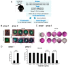
CLU seals the ocular surface barrier damaged by BAC. (A) One drop of a BAC solution (0.2% dissolved in PBS) was applied to the ocular surface twice (9 a.m., 5 p.m.) on day 1, and twice (9 a.m., 5 p.m.) on day 2. On the morning of day 3, the ocular surface was treated with a drop of CLU (100 µg/mL), or with vehicle alone (PBS). For comparison, unstressed eyes were similarly treated. Ten minutes after CLU treatment, eyes were enucleated and sealing was evaluated by in vitro rose bengal staining and imaging. (B) Images of enucleated eyes stained with rose bengal. The experiment was performed in duplicate (Set 1 and Set 2).
Figure 2
Figure 2
CLU seals the ocular surface barrier damaged by BAC in a concentration-dependent manner. (A) One drop of a BAC solution (0.2% dissolved in PBS) was applied to the ocular surface twice (9 a.m., 5 p.m.) on day 1, and twice (9 a.m., 5 p.m.) on day 2. In one group of mice (group 1), right eyes were subjected to the BAC stress protocol, while left eyes were left unstressed. In a second group of mice (group 2), both right and left eyes were subjected to the BAC stress protocol. On the morning of day 3, right eyes of the second group were treated topically with CLU (0.1, 1 or 10 µg/mL) and left eyes were treated with vehicle alone (PBS). Within 10 min, sealing was assayed in eyes of both groups by staining in situ with fluorescein, or by enucleating eyes and staining in vitro with rose bengal. Shown are representative eyes stained with (B) fluorescein or (C) rose bengal. (D) Quantification of rose bengal staining for all mice for which a representative example is shown in (C), expressed as the mean ± standard deviation (n = 3). Statistical significance was determined by paired T-test and p-values are indicated where significant.
Figure 3
Figure 3
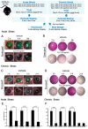
CLU sealing of the ocular surface barrier damaged by BAC persists for 4 to 6 h. Acute Stress. One drop of a BAC solution (0.2% dissolved in PBS) was applied to the ocular surface twice (9 a.m., 5 p.m.) on day 1, and twice (9 a.m., 5 p.m.) on day 2. On the morning of day 3, the ocular surface was treated topically with CLU (10 µg/mL) or PBS vehicle alone. Sealing was then assayed every two hours subsequently, for 6 h total. Shown are representative eyes stained with (A) fluorescein, or (B) rose bengal. Chronic Stress. One drop of a BAC solution (0.2% dissolved in PBS) was applied to the ocular surface twice (9 a.m., 5 p.m.) on day 1, and twice (9 a.m., 5 p.m.) on day 2. On the morning of day 3, BAC was applied one more time, then the ocular surface was treated topically with CLU (10 µg/mL) or PBS vehicle alone. Sealing was assayed every two hours subsequently for 6 h, then at 24 h. Shown are representative eyes stained with (C) fluorescein, or (D) rose bengal. Quantification. Rose bengal staining in (E) acute stress and (F) chronic stress experiments is quantified and expressed on the graphs shown as the mean ± SD (n = 3). Statistical significance was determined by paired T-test and p-values are indicated over the lines connecting bars which are significantly different.
Figure 4
Figure 4
Expression and characterization of GMP-compatible rhCLU. (A) CLU Structure. The secretory signal peptide is proteolytically cleaved from the precursor polypeptide chain and subsequently the chain is cleaved again between residues Arg227–Ser228 to generate an α-chain and a β-chain. These are assembled in anti-parallel fashion to generate a heterodimeric molecule in which the cysteine-rich centers (red boxes) are linked by five disulfide bonds (black rectangles) and flanked by five predicted amphipathic α-helices (yellow boxes). Amino acid numbering for the N- and C-termini, signal peptide, cleavage site, and predicted sites for N-linked glycosylation are indicated (white spots). (B) Processing and Purity. Shown is a Coomassie blue-stained SDS-PAGE. Lane 1: nonreduced pCLU; Lane 2: non-reduced rhCLU-αC-H2S; Lane 3: molecular size standard in kDa; Lane 4: pCLU reduced using β-mercaptoethanol; Lane 5: rhCLU-αC-H2S reduced using β-mercaptoethanol. (C) Identity. Shown are Western blots. Lanes 2, 5, 8: non-reduced pCLU; Lanes 3, 6, 9: Molecular size standard. The upper pink band is 75 kDa. Left Panel. CLU antibody probe. Middle Panel. Strep tag antibody probe. Right Panel. Hexahistidine tag antibody probe. Lanes 1, 4, 7: non-reduced rhCLU-αC-H2ST. (D) Molecular Chaperone Activity. The client protein, CS (citrate synthase), was heated to 43 °C to induce misfolding and aggregation. rhCLU-αC-H2S or pCLU were mixed with the client at two molar ratios: 1:1 or 2:1, client to chaperone. The negative control, bovine serum albumin (BSA), was assessed in parallel. Aggregation of the client was measured over a 200-min time course as absorbance at 360 nm.
Figure 5
Figure 5
Efficacy and selectivity of GMP-compatible rhCLU in sealing the ocular surface barrier damaged by BAC. (A) A single drop of a BAC solution (0.2% dissolved in PBS) was applied to the ocular surface twice (9 a.m., 5 p.m.) on day 1, and twice (9 a.m., 5 p.m.) on day 2. In one group of mice, right eyes were subjected to the 2-day BAC protocol, while left eyes were left unstressed. In a second group of mice, both right and left eyes were subjected to the 2-day BAC stress protocol. One the morning of day 3, right eyes of the second group were treated topically with rhCLU-αC-H2ST (rhCLU) formulated in PBS at 100 µg/mL or with one of the reference proteins, recombinant human LCN1 or recombinant human TIMP1, also formulated in PBS at 100 µg/mL; left eyes were treated with PBS vehicle alone. Within 10 min, sealing was assayed in eyes of both groups by staining with fluorescein (B) or rose bengal (C). Shown are representative examples from each subgroup. (D) Quantified values for rose bengal staining, expressed as the mean ± SD (n = 3). Statistical significance was determined by ANOVA and significant p-values are indicated.
Figure 6
Figure 6
The antioxidant capacity of CLU is low. Total antioxidant capacity for human pCLU and rhCLU-αC-H2ST (rhCLU) was compared using the OxiSelect™ Total Antioxidant Capacity Assay Kit (Cell Biolabs, Inc., San Diego, CA, USA), according to the manufacturer’s directions. Absorbance at 490 nM was determined with a Synergy H1 microplate reader at the beginning and end of the reaction, and the difference was calculated and plotted on the graph. Bovine serum ALB (BSA) and Trolox served as positive standards and glucose served as a negative standard.

References

    1. Gipson I.K. The ocular surface: The challenge to enable and protect vision: The Friedenwald lecture. Investig. Ophthalmol. Vis. Sci. 2007;48:4391–4398. doi: 10.1167/iovs.07-0770. - DOI - PMC - PubMed
    1. Hanna C., O’Brien J.E. Cell production and migration in the epithelial layer of the cornea. Arch. Ophthalmol. 1960;64:536–539. doi: 10.1001/archopht.1960.01840010538009. - DOI - PubMed
    1. Hanna C., Bicknell D.S., O’Brien J.E. Cell turnover in the adult human eye. Arch. Ophthalmol. 1961;65:695–698. doi: 10.1001/archopht.1961.01840020697016. - DOI - PubMed
    1. Ong H.S., Dart J.K. Managing ocular surface disease: A common-sense approach. Community Eye Health. 2016;29:44–46. - PMC - PubMed
    1. TFOS Report of the International Dry Eye Workshop (DEWS) Ocul. Surf. 2007;5:65–204. - PubMed