. 2023 Jan 7;24(2):1221.
doi: 10.3390/ijms24021221.

Estradiol and Estrone Have Different Biological Functions to Induce NF-κB-Driven Inflammation, EMT and Stemness in ER+ Cancer Cells

Affiliations

Estradiol and Estrone Have Different Biological Functions to Induce NF-κB-Driven Inflammation, EMT and Stemness in ER+ Cancer Cells

Ana Belén Diaz-Ruano et al. Int J Mol Sci. .

Abstract

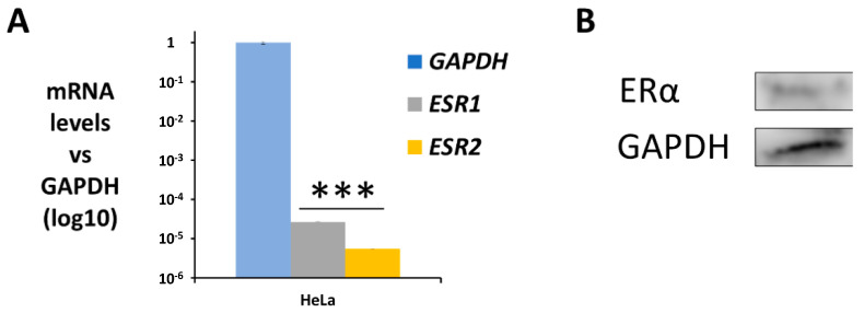
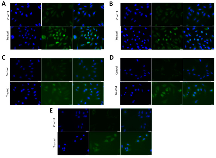
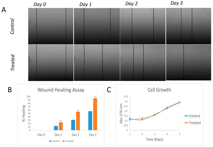
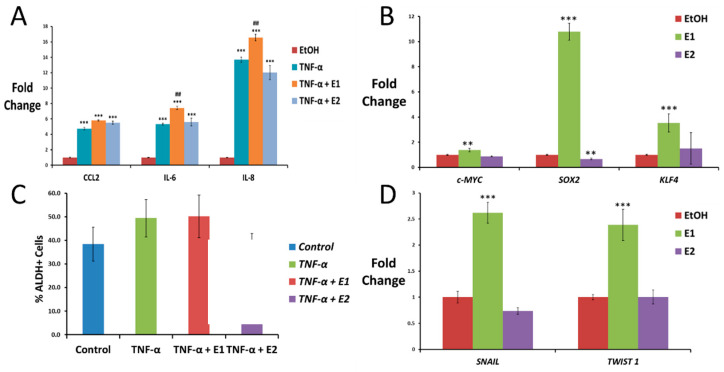
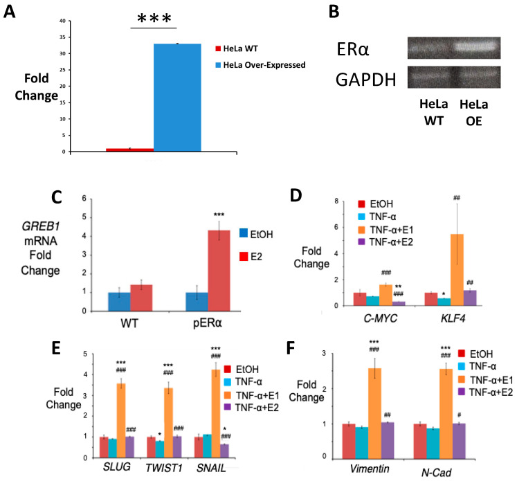
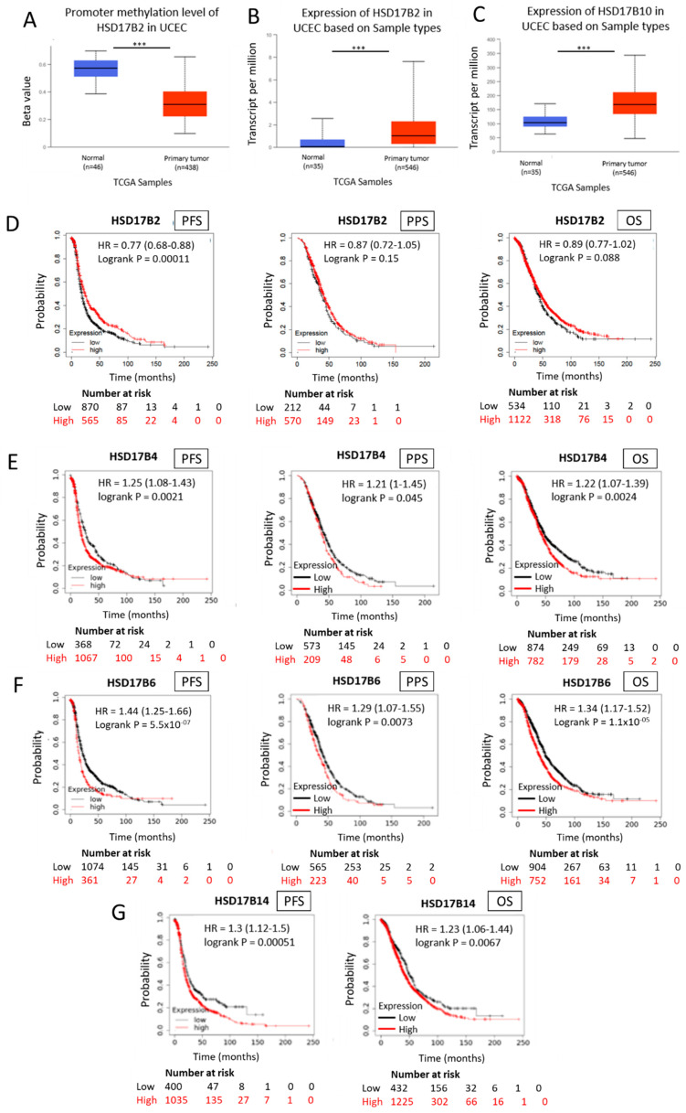
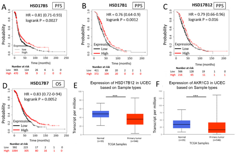
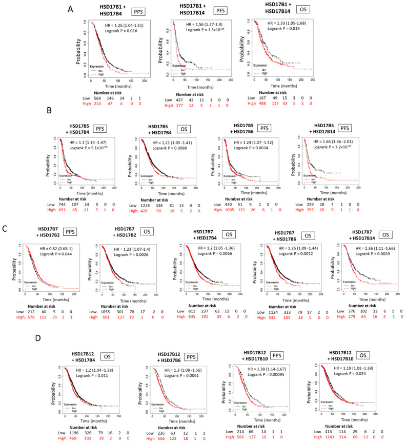
In general, the risk of being diagnosed with cancer increases with age; however, the development of estrogen-receptor-positive (ER+) cancer types in women are more closely related to menopausal status than age. In fact, the general risk factors for cancer development, such as obesity-induced inflammation, show differences in their association with ER+ cancer risk in pre- and postmenopausal women. Here, we tested the role of the principal estrogens in the bloodstream before and after menopause, estradiol (E2) and estrone (E1), respectively, on inflammation, epithelial-to-mesenchymal transition (EMT) and cancer stem cell enrichment in the human ER+ cervical cancer cell line HeLa. Our results demonstrate that E1, contrary to E2, is pro-inflammatory, increases embryonic stem-transcription factors (ES-TFs) expression and induces EMT in ER+ HeLa cells. Moreover, we observed that high intratumoural expression levels of 17β-Hydroxysteroid dehydrogenase (HSD17B) isoforms involved in E1 synthesis is a poor prognosis factor, while overexpression of E2-synthetizing HSD17B isoforms is associated with a better outcome, for patients diagnosed with ER+ ovarian and uterine corpus carcinomas. This work demonstrates that E1 and E2 have different biological functions in ER+ gynaecologic cancers. These results open a new line of research in the study of ER+ cancer subtypes, highlighting the potential key oncogenic role of E1 and HSD17B E1-synthesizing enzymes in the development and progression of these diseases.

Keywords: ER+ cancer; HSD17B; NF-κB; estradiol; estrone; inflammation.


Conflict of interest statement

The authors declare no conflict of interest.

Figures

Figure 1
Figure 1
ER expression in HeLa cells. (A) Gene expression profile of both ER isoforms using qPCR, graphed relative to the housekeeping GAPDH as mean ± SEM (n = 3). (B) Western Blot of ERα expression using GAPDH as housekeeping (*** p < 0.001).
Figure 2
Figure 2
NF-κB nuclear translocation using TNFα treatment in HeLa cells. (A–E) Fluorescent immunostaining of HeLa cells untreated (control) or treated with 10 ng/mL TNF-α for 45 min using antibodies against human RelA/p65 (A), RelB (B), cRel (C), p50 (D), p52 (E) (green). Nuclei were stained with DAPI (blue).
Figure 3
Figure 3
Wound-healing and proliferation assays in TNFα-treated in HeLa cells. Images of wound-healing assay performed on HeLa cells. Treated vs control (A) and graph representation (n = 3; *** p < 0.001 vs control) (B). Study of cell growth differences between treated and untreated cells (C).
Figure 4
Figure 4
Effect of TNF-α, estrone and estradiol on driving inflammation, stemness and EMT in HeLa cells. (A) Pro-inflammatory cytokine expression measured using qPCR in HeLa cells after treatment with 10 ng/mL TNF-α for 4 h alone or in combination with 10 nM E1 or E2 (data normalized to 1 for control (EtOH) using GAPDH as the housekeeping gene). (B) qPCR for the expression of the ES-TF c-MYC and SOX2 in HeLa cells after 3 weeks of exposure to 10 nM E1 or E2 (data normalized to 1 for control (EtOH) using GAPDH as the housekeeping gene). (C) Expression of ALDH activity measured using flow cytometry in HeLa cells untreated (control) or treated with 10 ng/mL TNF-α alone or in combination with 10 nM E1 or E2 for 3 weeks. (D) EMT transcription factor expression measured using qPCR in HeLa cells exposed to 10 nM E1 or E2 for 3 weeks (data normalized to 1 for control using GAPDH as the housekeeping gene). All data are graphed as mean ± SEM from experiments performed in triplicates and repeated at least 3 times. ** p < 0.01 *** p < 0.001 vs control; ## p < 0.05 vs TNF-α.
Figure 5
Figure 5
Effect of TNF-α, estrone and estradiol on driving stemness and EMT in HeLa pERα cells. (A) Western Blot of ERα expression using GAPDH as housekeeping. (B) Gene expression profile of ERα using qPCR, graphed as mean ± SEM (n = 3) using WT as normalizer and GAPDH as housekeeping gene (*** p < 0.001 vs Wild Type). (C) qPCR for the expression of GREB1 in WT and pERα HeLa cells treated with 10 nM E2 for 4 h (data normalized to 1 for control (EtOH) using GAPDH as the housekeeping gene). (D) ES-TF expression in HeLa pERα cells assayed using qPCR after exposure to 10 ng/mL TNF-α alone or in combination with 10 nM E1 or E2 for at least 1 week (data normalized to 1 for control using GAPDH as the housekeeping gene). (C,D) qPCR analysis for the expression of the EMT transcription factors SLUG, TWIST1 and SNAIL (E) and the EMT markers Vimentin and N-Cadherin (F) in HeLa pERα cells treated with the vehicle (EtOH, control) or 10 ng/mL TNF-α alone or in combination with 10 nM E1 or E2 for 3 weeks (data normalized to 1 for control using GAPDH as the housekeeping gene). All data are graphed as mean ± SEM from experiments performed in triplicates and repeated at least 3 times. * p < 0.05 ** p < 0.01 *** p < 0.001 vs control; # p < 0.05 ## p < 0.01 ### p < 0.001 vs TNF-α.
Figure 6
Figure 6
Expression and methylation of HSD17B enzymes involved in estrone synthesis and ovarian cancer patient outcome (BF). Methylation levels of the HSD17B2 enzyme in normal and tumour tissue samples obtained from “The Cancer Genome Atlas” (TCGA) database (A). Expression levels of the HSD17B2 enzyme in normal and tumour tissue samples obtained from the TCGA (B). Expression levels of the HSD17B10 enzyme in normal and tumour tissue samples obtained from the TCGA (C). Kaplan–Meier curves comparing low (black line) vs high (red line) tumoural expression levels of HSD17B2, HSDD17B4 and HSD17B6 enzymes involved in E1 synthesis and progression-free survival (PFS), post-progression-free survival (PPS) and overall survival (OS) in ovarian cancer patients (HR = Hazard Ratio) (DF) in the case of HSD17B14 only PFS and OS are represented (G). *** p < 0.001.
Figure 7
Figure 7
Expression of HSD17B enzymes involved in estradiol synthesis and ovarian cancer patient outcome (AD). Kaplan–Meier curves comparing low (black line) vs high (red line) tumour expression levels of different enzymes of the HSD17B family; HSD17B5 enzyme in PFS (A); HSD17B1 enzyme in PPS (B); HSD17B12 enzyme in PPS (C); HSD17B7 enzyme in OS (D). Expression of HSD17B12 in normal and tumour samples taken from the TCGA program (E). Expression of AKR1C3 in normal and tumour samples taken from the TCGA program (F). *** p < 0.001.
Figure 8
Figure 8
Ovarian cancer patient outcome based on combined low and high expression of E2- and E1-synthetizing HSD17B enzymes, respectively. (AD) Kaplan–Meier curves comparing the correlation between low intratumoural expression levels of the E2-synthetizing enzymes HSD17B1 (A), HSD17B5 (B), HSD17B7 (C) and HSD17B12 (D); combined with high intratumorual expression of the different E1-synthetizing HSD17B isoforms indicated, including HSD17B2, HSD17B4, HSD17B6, HSD17B10 and HSD17B14; and PPS, PFS and OS in ovarian cancer patients.

References

    1. Bhardwaj P., Au C.C., Benito-Martin A., Ladumor H., Oshchepkova S., Moges R., Brown K.A. Estrogens and breast cancer: Mechanisms involved in obesity-related development, growth and progression. J. Steroid Biochem. Mol. Biol. 2019;189:161–170. doi: 10.1016/j.jsbmb.2019.03.002. - DOI - PMC - PubMed
    1. Deroo B.J., Korach K.S. Estrogen receptors and human disease. J. Clin. Investig. 2006;116:561–570. doi: 10.1172/JCI27987. - DOI - PMC - PubMed
    1. Richardson H., Ho V., Pasquet R., Singh R.J., Goetz M.P., Tu D., Goss P.E., Ingle J.N. Baseline estrogen levels in postmenopausal women participating in the MAP.3 breast cancer chemoprevention trial. Menopause. 2020;27:693–700. doi: 10.1097/GME.0000000000001568. - DOI - PMC - PubMed
    1. Cui J., Shen Y., Li R. Estrogen synthesis and signaling pathways during aging: From periphery to brain. Trends Mol. Med. 2013;19:197–209. doi: 10.1016/j.molmed.2012.12.007. - DOI - PMC - PubMed
    1. Ferlay J., Soerjomataram I., Dikshit R., Eser S., Mathers C., Rebelo M., Parkin D.M., Forman D., Bray F. Cancer incidence and mortality worldwide: Sources, methods and major patterns in GLOBOCAN 2012. Int. J. Cancer. 2015;136:E359–E386. doi: 10.1002/ijc.29210. - DOI - PubMed