. 2023 Jan 8;24(2):1241.
doi: 10.3390/ijms24021241.

Mitogen-like Cerium-Based Nanoparticles Protect Schmidtea mediterranea against Severe Doses of X-rays

Affiliations

Mitogen-like Cerium-Based Nanoparticles Protect Schmidtea mediterranea against Severe Doses of X-rays

Kristina O Filippova et al. Int J Mol Sci. .

Abstract

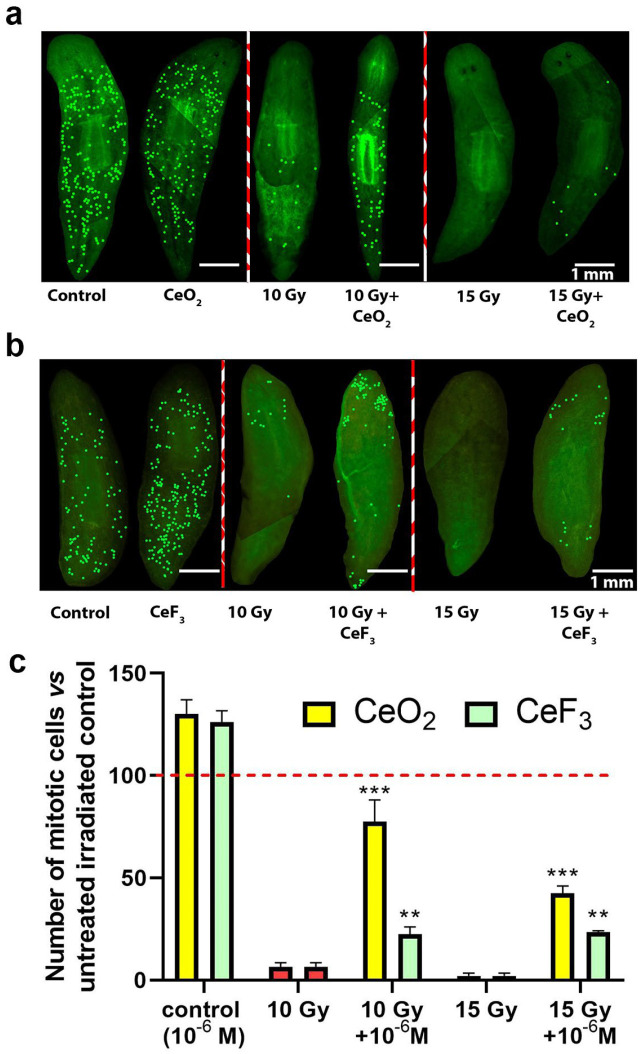
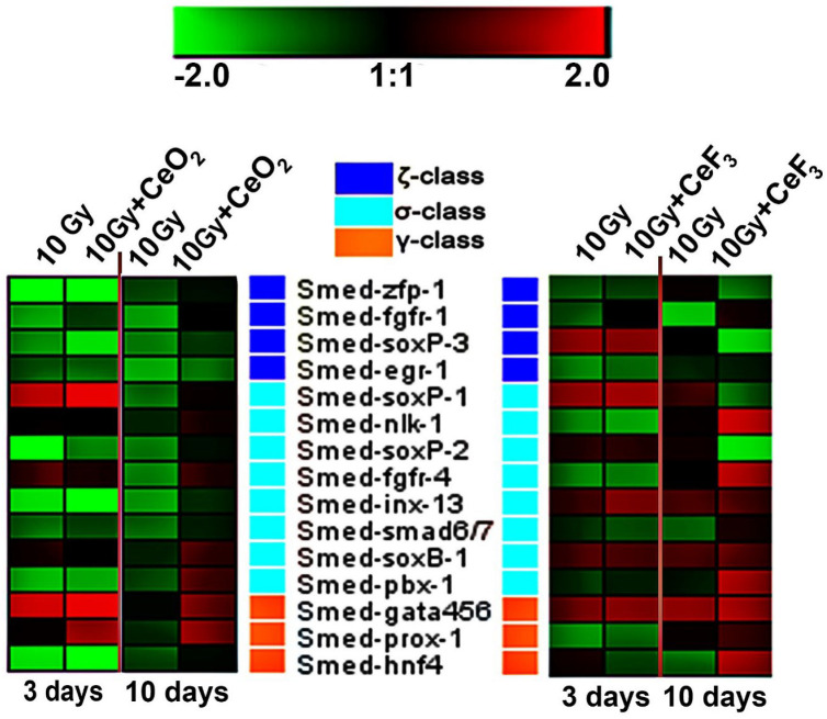
Novel radioprotectors are strongly demanded due to their numerous applications in radiobiology and biomedicine, e.g., for facilitating the remedy after cancer radiotherapy. Currently, cerium-containing nanomaterials are regarded as promising inorganic radioprotectors due to their unrivaled antioxidant activity based on their ability to mimic the action of natural redox enzymes like catalase and superoxide dismutase and to neutralize reactive oxygen species (ROS), which are by far the main damaging factors of ionizing radiation. The freshwater planarian flatworms are considered a promising system for testing new radioprotectors, due to the high regenerative potential of these species and an excessive amount of proliferating stem cells (neoblasts) in their bodies. Using planarian Schmidtea mediterranea, we tested CeO2 nanoparticles, well known for their antioxidant activity, along with much less studied CeF3 nanoparticles, for their radioprotective potential. In addition, both CeO2 and CeF3 nanoparticles improve planarian head blastema regeneration after ionizing irradiation by enhancing blastema growth, increasing the number of mitoses and neoblasts' survival, and modulating the expression of genes responsible for the proliferation and differentiation of neoblasts. The CeO2 nanoparticles' action stems directly from their redox activity as ROS scavengers, while the CeF3 nanoparticles' action is mediated by overexpression of "wound-induced genes" and neoblast- and stem cell-regulating genes.

Keywords: X-ray; cerium fluoride nanoparticles; cerium oxide nanoparticles; planarians; radioprotection.


Conflict of interest statement

The authors declare no conflict of interest.

Figures

Figure 1
Figure 1
Transmission electron microscopy (a,b), dynamic light scattering (c,d) in water, and UV absorption spectra (e,f) of cerium oxide (CeO2) and cerium fluoride (CeF3) nanoparticles. Insets in (a,b) show selected-area electron diffraction data.
Figure 2
Figure 2
Concentration dependencies of hydrogen peroxide formation induced by X-ray irradiation (1 Gy) in a buffer solution containing CeF3 and CeO2 NPs. The mean values of three independent experiments and their standard errors are given.
Figure 3
Figure 3
Radioprotective effect of CeO2 nanoparticles on regenerating planarian blastemas after X-ray irradiation (10 Gy and 15 Gy). Brown to light-brown bars represent the percentage of blastema regeneration rate; the regeneration rate of non-irradiated animals was taken as 100% (red line). * p < 0.05 (difference from the control group). M ± SD, n = 90. IR—X-ray irradiation.
Figure 4
Figure 4
Radioprotective effect of CeF3 nanoparticles on regenerating planarian blastema after irradiation with 10 Gy and 15 Gy. Green to light-green bars represent the percentage of blastema regeneration rate; the regeneration rate of non-irradiated animals was taken as 100% (red line). * p < 0.05 (difference from the control group). M ± SD, n = 90. IR—X-ray irradiation.
Figure 5
Figure 5
Mitotic activity of regenerating planarians pretreated with CeO2 and CeF3 nanoparticles and X-ray irradiated. Photographs of immunostained planarians after X-rays irradiation with CeO2 (a) and cerium fluoride (b) nanoparticles. Quantitative analysis of the level of mitotic cells (c). This concentration of nanoparticles (106 M) was chosen as it gave the most pronounced effect. Significance was estimated by one-way analysis of variance (one-way ANOVA). Data are presented as means. Significant statistical difference ** p < 0.001, *** p < 0.05 compared to the untreated control (indicated by the red line).
Figure 6
Figure 6
Expression of 3 classes of neoblast marker genes in regenerating planarians on days 3 and 10 after 10 Gy X-ray irradiation. The intensity scale of the standardized expression values ranges from −2 (green: low expression) to +2 (red: high expression), with a 1:1 intensity value (black) representing the control (non-treated). The data in the heat maps are from the non-irradiated control group. A non-irradiated control group without CeO2 or CeF3 nanoparticle pretreatment was taken as a control.

References

    1. Reisz J.A., Bansal N., Qian J., Zhao W., Furdui C.M. Effects of ionizing radiation on biological molecules—Mechanisms of damage and emerging methods of detection. Antioxid. Redox Signal. 2014;21:260–292. doi: 10.1089/ars.2013.5489. - DOI - PMC - PubMed
    1. Cadet J., Douki T., Gasparutto D., Ravanat J.L. Oxidative damage to DNA: Formation, measurement and biochemical features. Mutat. Res. 2003;531:5–23. - PubMed
    1. Von Sonntag C. Nucleobases, Nucleosides and Nucleotides. Free-Radical-Induced DNA Damage and Its Repair: A Chemical Perspective. Springer; Berlin/Heidelberg, Germany: 2006. pp. 211–334.
    1. Azzam E.I., Jay-Gerin J.P., Pain D. Ionizing radiation-induced metabolic oxidative stress and prolonged cell injury. Cancer Lett. 2012;327:48–60. doi: 10.1016/j.canlet.2011.12.012. - DOI - PMC - PubMed
    1. Mishra K., Alsbeih G. Appraisal of biochemical classes of radioprotectors: Evidence, current status and guidelines for future development. Biotech. 2017;7:292. doi: 10.1007/s13205-017-0925-0. - DOI - PMC - PubMed

LinkOut - more resources