Review
. 2023 Jan 10;24(2):1353.
doi: 10.3390/ijms24021353.

The Phospholipase A2 Superfamily: Structure, Isozymes, Catalysis, Physiologic and Pathologic Roles

Affiliations
Review

The Phospholipase A2 Superfamily: Structure, Isozymes, Catalysis, Physiologic and Pathologic Roles

Shibbir Ahmed Khan et al. Int J Mol Sci. .

Abstract

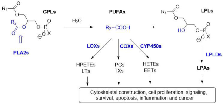
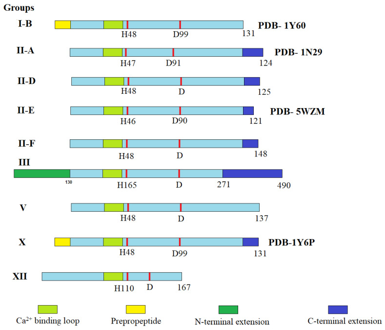
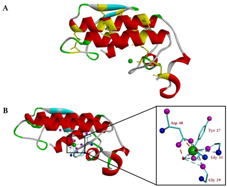
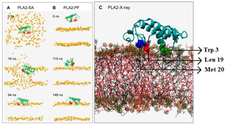
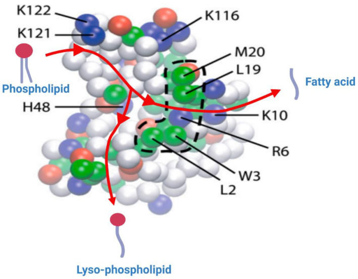
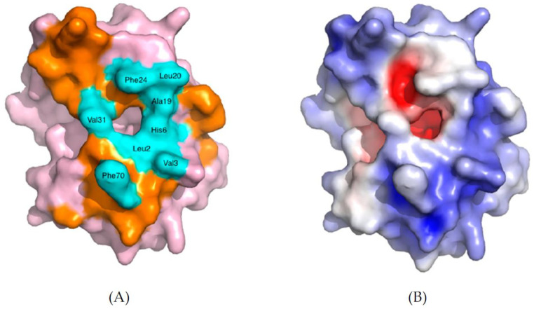
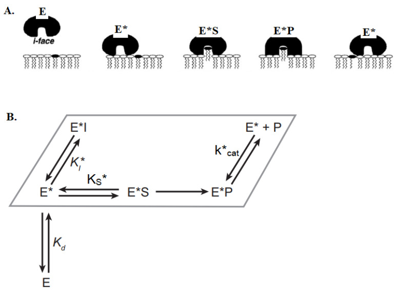
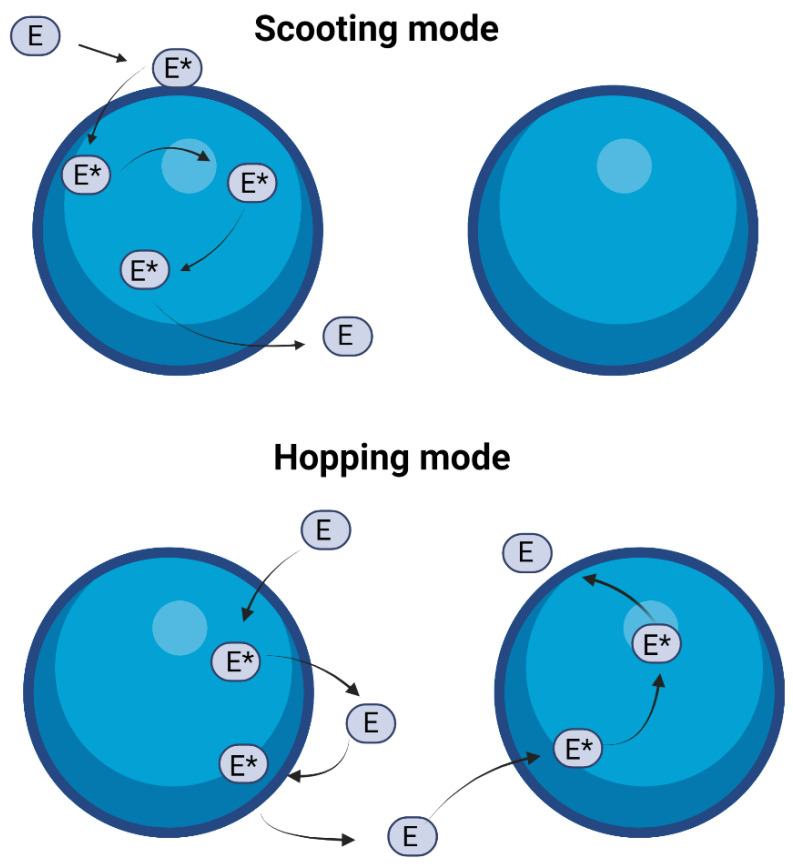
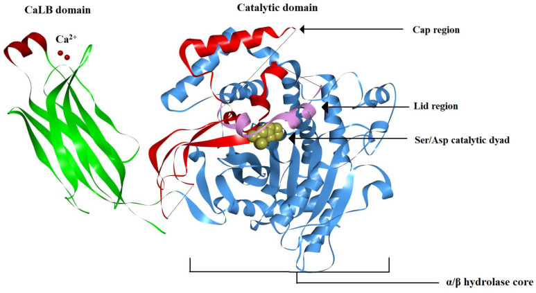
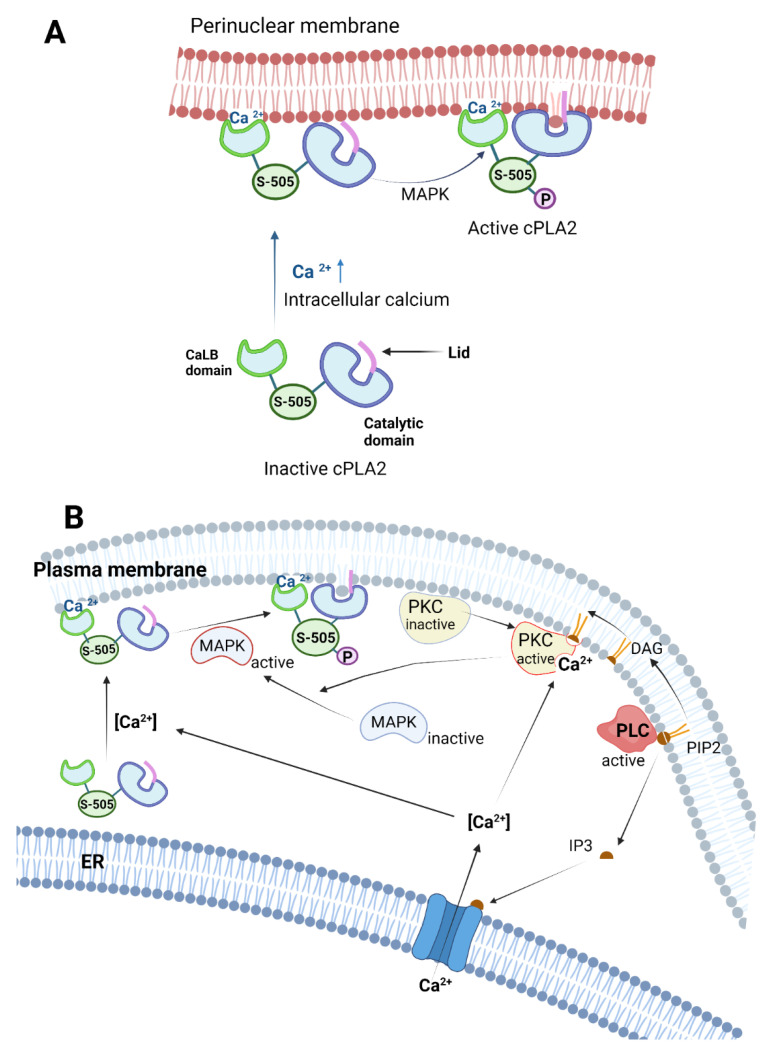
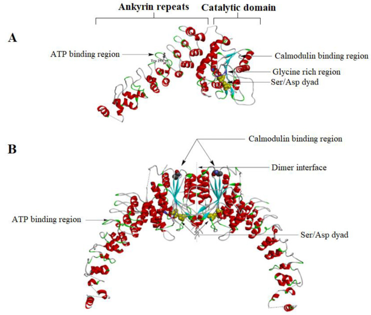
The phospholipase A2 (PLA2) superfamily of phospholipase enzymes hydrolyzes the ester bond at the sn-2 position of the phospholipids, generating a free fatty acid and a lysophospholipid. The PLA2s are amphiphilic in nature and work only at the water/lipid interface, acting on phospholipid assemblies rather than on isolated single phospholipids. The superfamily of PLA2 comprises at least six big families of isoenzymes, based on their structure, location, substrate specificity and physiologic roles. We are reviewing the secreted PLA2 (sPLA2), cytosolic PLA2 (cPLA2), Ca2+-independent PLA2 (iPLA2), lipoprotein-associated PLA2 (LpPLA2), lysosomal PLA2 (LPLA2) and adipose-tissue-specific PLA2 (AdPLA2), focusing on the differences in their structure, mechanism of action, substrate specificity, interfacial kinetics and tissue distribution. The PLA2s play important roles both physiologically and pathologically, with their expression increasing significantly in diseases such as sepsis, inflammation, different cancers, glaucoma, obesity and Alzheimer's disease, which are also detailed in this review.

Keywords: activation mechanism; catalysis; isozyme; kinetics; pathology; phospholipase A2; physiology; structure; subcellular localization; substrate specificity; tissue distribution.


Conflict of interest statement

The authors declare no conflict of interest.

Figures

Figure 1
Figure 1
Schematic diagram of the fate of the phospholipids catalyzed by the PLA2s and further transformation into inflammatory mediators: PLA2s selectively hydrolyze the ester bond in sn-2 position of glycerophospholipids (GPLs) and release lysophospholipids (LPLs) and free fatty acids—mostly polyunsaturated fatty acids (PUFAs). PUFAs such as AA are metabolized by cyclooxygenases (COXs), lipoxygenases (LOXs) and cytochromes p450 (CYP450s) metabolic pathways into eicosanoids such as prostaglandins (PGs), thromboxanes (TXs), hydroperoxy-eicosatetraenoic acids (HPETEs), leukotrienes (LTs), hydroxy-eicosatetraenoic acids (HETEs) and epoxyeicosatrienoic acids (EETs). The LPLs can be further hydrolyzed by lysophospholipases D (LPLDs) into lysophosphatidic acids (LPAs) [3].
Figure 2
Figure 2
Schematic presentation of the different groups of secreted PLA2s found in humans (adapted from [6]). The Ca2+-binding loop is shown in light green, the key active site residues (His and Asp) are shown as red bars, and N-terminal (deep green) and C- terminal (deep blue) extension residues are also highlighted. Adapted with permission from [6]. Copyright 2011, American Chemical Society.
Figure 3
Figure 3
(A) Top view of the crystal structure of G-IA sPLA2 (Naja naja) (PDB-1PSH). The α-helices and β-sheets are shown in red and light blue. The cysteines forming the seven disulfide bonds are shown in yellow, also marking in yellow their position on the protein backbone. The calcium ion is shown as a green sphere in the enzyme active site. (B) Side view of the crystal structure of the sPLA2 with the Ca2+ coordination shown in detail in the zoomed insert. The oxygen atoms and the nitrogen atoms of the amino acids coordinated with Ca2+ (green sphere) are colored pink and blue, respectively. The Ca2+ is directly coordinated with carboxy group from Asp48 of one α-helix and by the C=O backbone groups of Tyr27, Gly29 and Gly31 from the opposite loop. (Image generated using BIOVIA Discovery studio.)
Figure 4
Figure 4
Proposed catalytic mechanism for sPLA2. Asp98 and His47 constitute the catalytic dyad which plays a key role in catalysis. These two residues are hydrogen-bonded together and further connected with the Ca2+ ion with the help of two water molecules (w5 and w6). His47 facilitates the deprotonation of w5 water molecule, subsequently attacks the sn-2 carbonyl group of the substrate and forms the tetrahedral intermediate. The rate-limiting step is the decomposition of the tetrahedral intermediate [20].
Figure 5
Figure 5
Cartoon depicting the X-ray crystal structure of cobra venom sPLA2 (wireframe) with dimyristoyl phosphatidylethanolamine (shown in a space-filling model) bound to it. The active site dyad His48/Asp99 and the Ca2+ ion are depicted together as a purple sphere. The aromatic interfacial residues Phe-5, Trp-19, Tyr-52 and Tyr-69 of PLA2 are shown in white. These residues of the enzyme are reaching as deep as 9–10 carbons in the acyl chain of the fatty acid while the rest of the chain is submerged into the lipid bilayer. Reprinted with permission from [21]. Copyright 1994, Elsevier.
Figure 6
Figure 6
Snapshots from the CG-MD simulations at different timeframes: (A) sPLA2-SA (self-assembled simulation); (B) sPLA2-PF (preformed simulation). The backbone of the enzyme is shown in green; the hydrophobic anchoring residues are shown in red; and the active site residue (H48) is depicted as a blue sphere; (C) snapshot of atomistic MD simulation of sPLA2 (PLA2-X-ray) on phospholipid bilayer. The protein backbone is depicted in light blue, and the three hydrophobic residues Trp-3, Leu-19 and Met-20 are colored in green, red, and deep blue, respectively, in a space-filling model. Reprinted/adapted with permission from [22]. Copyright 2008, Elsevier.
Figure 7
Figure 7
Representation of the bottom (inter)face of the sPLA2, which remains in contact with the lipid bilayer, generated through the CG simulation. The amino acid residues which remained deep in the bilayer are represented in strong colors, and the ones that are away from the bilayer are shown in faded colors. The hydrophobic, basic, acidic and polar residues are shown as green, blue, red and white spheres, respectively. The broken line delimiting the cluster of hydrophobic residues (Trp-3, Leu-19, Met-20) highlights the key residues believed to anchor the sPLA2 into the lipid bilayer. The basic residues (Arg-6, Lys-10, Lys-116, Lys-121 and Lys-122) form electrostatic interactions with the phosphate group and with the carbonyl group of the lipid substrate. Reprinted/adapted with permission from [22]. Copyright 2008, Elsevier.
Figure 8
Figure 8
Space-filling model of human group IIA sPLA2 (PDB—3U8B). (A) The amino acids which form the i-face are colored orange, and the ones which create the entrance are highlighted in cyan blue, while the rest of the protein is shown in pink. (B) The electrostatic charge distribution is depicted either in blue (positive charge) or in red (negative charge). The white areas charge neutrality (non-polar), which corresponds to the hydrophobic entrance of the left figure. Reprinted from [26].
Figure 9
Figure 9
Schematic representation of interfacial binding and catalytic action of sPLA2. (A) The binding of the sPLA2 at the lipid/water interface through the i-face is essential for catalysis. The enzyme passes along the horizontal plane on the bilayer, while hydrolyzing the phospholipid molecules and releasing the LPL and FA products [20]. (B) The minimal kinetic scheme for the catalytic cycle of sPLA2 is shown in a parallelogram box that represents the lipid bilayer. Here, E denotes enzyme in the aqueous phase, E* denotes enzyme in a membrane-bound state without substrate and Kd is the interfacial dissociation constant for the enzyme at the interface. KS, Kcat and KI are the dissociation constants for substrate, product and inhibitor, respectively. E*S and E*P denote the enzyme-bound substrate and enzyme-bound product, respectively. Adapted with permission from [20]. Copyright 2001, American Chemical Society.
Figure 10
Figure 10
Cartoon showing the two main modes in which sPLA2 enzyme may interact with the lipid vesicles. In scooting mode (top), the enzyme does not exchange vesicles after each catalytic turnover, and in hopping mode (bottom), the enzyme leaves the interface (of the vesicle) after each catalytic cycle and moves to another vesicle. Adapted with permission from [20]. Copyright 2001, American Chemical Society. Created with BioRender.com.
Figure 11
Figure 11
Ribbon diagram of a typical cPLA2 (PDB: 1CJY). The Ca2+-dependent lipid-binding (CaLB) domain is shown in green on the left side of the figure, with the two bound Ca2+ ions (required for protein translocation to the membrane) shown as red spheres. The catalytic domain is shown on the right side, the α/β hydrolase core is shown in blue and the catalytic dyad Ser/Asp is shown in yellow. The enzyme cap is shown in red, and the lid region covering the active site funnel is colored pink. The lid region prevents the lipid substrates to come in contact with the active site [31]. (Image generated using BIOVIA Discovery studio.)
Figure 12
Figure 12
Multiple activation pathways for cPLA2 via calcium, phosphorylation and secondary messengers. (A) Ca2+ ion helps in binding the CaLB domain of the cPLA2 with the membrane; however, the catalytic domain is not oriented in the right direction for hydrolyzing the phospholipid substrate. Phosphorylation in the flexible linker of the cPLA2 via MAPK induces optimal conformation of the catalytic domain, which facilitates partial penetration of the catalytic domain into the membrane. (B) The schematics for multiple regulation pathways for cPLA2. PIP2 is hydrolyzed by PLC to form secondary messengers such as IP3 and DAG. IP3 opens up the Ca2+ ion channel in the perinuclear membrane of the ER, which increases the intracellular Ca2+ concentration. The DAG and the Ca2+ simultaneously activates PKC, which subsequently activates MAPK. The activated MAPK phosphorylates the cPLA2, which further enhances the cPLA2 activity. Created with BioRender.com.
Figure 13
Figure 13
(A) The crystal structure of the monomeric form of iPLA2β (PDB- 6AUN). The ankyrin repeats (ANK) are shown on the left side and the catalytic domain on the right side. The linker between the ANK and the CAT (connected by a dashed line) is unresolved due to less electron density. Near the 6th ankyrin repeats, there is the Trp293 residue which is believed to be the ATP binding site. The catalytic dyad (Ser465 and Asp598) is shown in purple in the catalytic domain. The glycine-rich region, which forms a rigid handle, is shown in blue. Earlier studies showed it as the ATP binding domain. The CaM binding region is near residue 630. (B) The dimeric structure of the iPLA2. One CaM molecule binds two iPLA2 molecules and stabilizes the close confirmation of the active sites of the enzyme. Note that the two active sites are located near the dimeric interface (Image generated using BIOVIA Discovery studio).
Figure 14
Figure 14
The proposed common mechanism of cPLA2 and iPLA2 for hydrolysis of phospholipids. The two classes of PLA2s share a common Ser/Asp dyad.
Figure 15
Figure 15
Proposed schematic models of interfacial binding of four different families of PLA2s. HDX-MS technique was used to study the interaction of monomers of different PLA2s bound to membranes, using crystal structures of (A) group IA sPLA2 (PDB- 1PSH), (B) group IV cPLA2 (PDB- 1CJY), (C) group VIA iPLA2 (homology model based on lipase patatin structure, PDB- 1OXW) and (D) group VIIA Lp-PLA2 (PBD- 3D5E). Reprinted from [59].
Figure 16
Figure 16
Physiologic role of phospholipases in GI tract. Pancreatic sPLA2-IB is secreted in pancreatic juice as a zymogen and activated by trypsin or plasmin in the intestinal lumen. sPLA2-X is secreted by the enterocytes of the intestinal lumen. Both pancreatic sPLA2-IB and enterocytic sPLA2-X hydrolyze dietary and bile-secreted phospholipids. Phospholipase B is secreted in the distal intestine and hydrolyzes the undigested phospholipids yielding glycerylphosphocholine and FAs. Created with BioRender.com.
Figure 17
Figure 17
Physiological roles of sPLA2-IIA, as a key player in host defense against foreign self-assembled lipid structures in the blood, through the degradation of bacterial membranes, and in inflammation, where it hydrolyzes arachidonic acid from extracellular micro-vesicles, which subsequently feeds the inflammatory pathway. It also cleaves cardiolipin from extracellular mitochondria (released from activated neutrophils, platelets, mast cells, lymphocytes, etc.), liberating mitochondrial DNA that potentiates inflammation [62].

References

    1. Murakami M., Kudo I. Phospholipase A2. J. Biochem. 2002;131:285–292. doi: 10.1093/oxfordjournals.jbchem.a003101. - DOI - PubMed
    1. Cathcart M.-C., Lysaght J., Pidgeon G.P. Eicosanoid Signalling Pathways in the Development and Progression of Colorectal Cancer: Novel Approaches for Prevention/Intervention. Cancer Metastasis Rev. 2011;30:363–385. doi: 10.1007/s10555-011-9324-x. - DOI - PubMed
    1. Peng Z., Chang Y., Fan J., Ji W., Su C. Phospholipase A2 Superfamily in Cancer. Cancer Lett. 2021;497:165–177. doi: 10.1016/j.canlet.2020.10.021. - DOI - PubMed
    1. Moolenaar W.H., van Meeteren L.A., Giepmans B.N.G. The Ins and Outs of Lysophosphatidic Acid Signaling. BioEssays. 2004;26:870–881. doi: 10.1002/bies.20081. - DOI - PubMed
    1. Lam L., Ilies M.A. Evaluation of the Impact of Esterases and Lipases from the Circulatory System against Substrates of Different Lipophilicity. Int. J. Mol. Sci. 2022;23:1262. doi: 10.3390/ijms23031262. - DOI - PMC - PubMed