

First Clinical Report of the Intraoperative Macro- and Micro-Photodiagnosis and Photodynamic Therapy Using Talaporfin Sodium for a Patient with Disseminated Lumbar Medulloblastoma
- PMID: 36675360
- PMCID: PMC9867022
- DOI: 10.3390/jcm12020432
First Clinical Report of the Intraoperative Macro- and Micro-Photodiagnosis and Photodynamic Therapy Using Talaporfin Sodium for a Patient with Disseminated Lumbar Medulloblastoma
Abstract
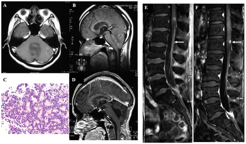
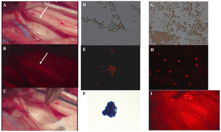
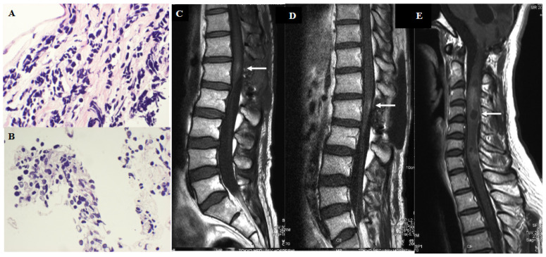
Photodiagnosis (PD) and photodynamic therapy (PDT) using the second-generation photosensitizer talaporfin sodium together with an exciting laser for primary intracranial malignant tumors is well recognized in Japan, and many medical institutions are introducing this new therapeutic option. In particular, intraoperative PDT using talaporfin sodium for infiltrating tumor cells in the cavity walls after the resection of malignant glioma is now covered by health insurance after receiving governmental approvement, and this method has been recommended in therapeutic guidelines for primary malignant brain tumors in Japan. On the other hand, experimental and clinical studies on the development of novel therapeutic strategies for malignant spinal cord tumors have not been reported to date, although their histological features are almost identical to those of intracranial malignant tumors. Therefore, the clinical outcomes of malignant spinal cord tumors have been less favorable than those of malignant brain tumors. In this report, we performed the PD and PDT using talaporfin sodium on a patient with a metastatic lumbar lesion that was detected on magnetic resonance image (MRI) 50 months after the resection of cerebellar medulloblastoma who presented with lumbago and sciatica. We were able to detect the target lesion in the conus medullaris using a surgical microscope, and detected the disseminated medulloblastoma cells floating in the cerebrospinal fluid using a compact fluorescence microscope. Furthermore, we performed PDT to the resected lumbar lesion with the adjuvant platinum-based chemotherapy, and the patient survived a meaningful life for more than 2 years after the lumbar surgery. This report describes the first case of a human patient in whom the efficacy of PD and PDT was demonstrated for a malignant spinal cord tumor.
Keywords: malignant spinal cord tumor; photodiagnosis; photodynamic therapy; talaporfin sodium.
Conflict of interest statement
This study is supported by Japan Agency for Medical Research and Development (Project number: 22ck0106738h0001).
Figures
Similar articles
-
Intraoperative Photodiagnosis for Malignant Glioma Using Photosensitizer Talaporfin Sodium.Front Surg. 2019 Mar 21;6:12. doi: 10.3389/fsurg.2019.00012. eCollection 2019. Front Surg. 2019. PMID: 30949484 Free PMC article.
-
Phase II clinical study on intraoperative photodynamic therapy with talaporfin sodium and semiconductor laser in patients with malignant brain tumors.J Neurosurg. 2013 Oct;119(4):845-52. doi: 10.3171/2013.7.JNS13415. Epub 2013 Aug 16. J Neurosurg. 2013. PMID: 23952800 Clinical Trial.
-
Preliminary clinical report on safety and efficacy of photodynamic therapy using talaporfin sodium for malignant gliomas.Photodiagnosis Photodyn Ther. 2012 Jun;9(2):91-9. doi: 10.1016/j.pdpdt.2012.01.001. Epub 2012 Feb 28. Photodiagnosis Photodyn Ther. 2012. PMID: 22594978 Clinical Trial.
-
Clinical Practice of Photodynamic Therapy Using Talaporfin Sodium for Esophageal Cancer.J Clin Med. 2021 Jun 24;10(13):2785. doi: 10.3390/jcm10132785. J Clin Med. 2021. PMID: 34202917 Free PMC article. Review.
-
Photodynamic Therapy for Malignant Brain Tumors.Neurol Med Chir (Tokyo). 2016;56(4):151-7. doi: 10.2176/nmc.ra.2015-0296. Epub 2016 Feb 16. Neurol Med Chir (Tokyo). 2016. PMID: 26888042 Free PMC article. Review.
Cited by
-
Smart molecular designs and applications of activatable organic photosensitizers.Nat Rev Chem. 2025 Jan;9(1):46-60. doi: 10.1038/s41570-024-00662-7. Epub 2024 Nov 6. Nat Rev Chem. 2025. PMID: 39506088 Review.
-
Combined use of 5-ALA-induced protoporphyrin IX and chlorin e6 for fluorescence diagnostics and photodynamic therapy of skin tumors.Lasers Med Sci. 2024 Oct 31;39(1):266. doi: 10.1007/s10103-024-04219-4. Lasers Med Sci. 2024. PMID: 39477891
References
-
- Muragaki Y., Akimoto J., Maruyama T., Iseki H., Ikuta S., Nitta M., Maebayashi K., Saito T., Okada Y., Kaneko S., et al. Phase II clinical study on intraoperative photodynamic therapy with talaporfin sodium and semiconductor laser. J. Neurosurg. 2013;119:845–852. doi: 10.3171/2013.7.JNS13415. - DOI - PubMed
-
- Nitta M., Muragaki Y., Maruyama T., Iseki H., Komori T., Ikuta S., Saito T., Yasuda T., Hosono J., Okamoto S., et al. Role of photodynamic therapy using talaporfin sodium and a semiconductor laser in patients with newly diagnoses glioblastoma. J. Neurosurg. 2018;12:1–8. doi: 10.3171/2018.7.JNS18422. - DOI - PubMed
-
- Kobayashi T., Nitta M., Shimizu K., Saito T., Tsuzuki S., Fukui A., Koriyama S., Kuwano A., Komori T., Masui K., et al. Therapeutic options for recurrent glioblastoma- Efficacy of talaporfin sodium mediated photodynamic therapy. Pharmaceutics. 2022;14:353. doi: 10.3390/pharmaceutics14020353. - DOI - PMC - PubMed
LinkOut - more resources
Full Text Sources
