

Corneal Sub-Basal Nerve Plexus in Non-Diabetic Small Fiber Polyneuropathies and the Diagnostic Role of In Vivo Corneal Confocal Microscopy
- PMID: 36675593
- PMCID: PMC9862881
- DOI: 10.3390/jcm12020664
Corneal Sub-Basal Nerve Plexus in Non-Diabetic Small Fiber Polyneuropathies and the Diagnostic Role of In Vivo Corneal Confocal Microscopy
Abstract
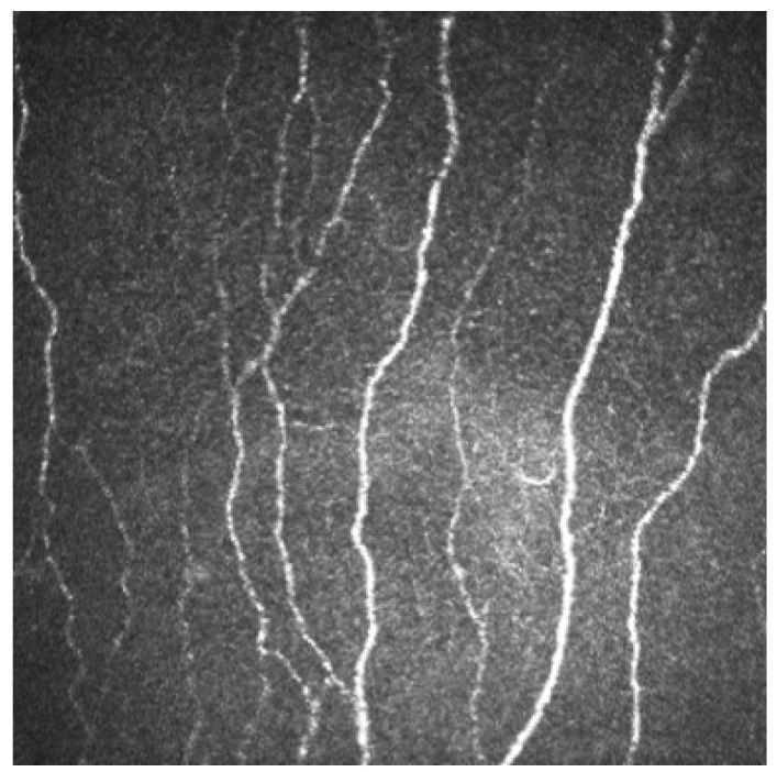
In vivo corneal confocal microscopy (IVCM) allows the immediate analysis of the corneal nerve quantity and morphology. This method became, an indispensable tool for the tropism examination, as it evaluates the small fiber plexus in the cornea. The IVCM provides us with direct information on the health of the sub-basal nerve plexus and indirectly on the peripheral nerve status. It is an important tool used to investigate peripheral polyneuropathies. Small-fiber neuropathy (SFN) is a group of neurological disorders characterized by neuropathic pain symptoms and autonomic complaints due to the selective involvement of thinly myelinated Aδ-fibers and unmyelinated C-fibers. Accurate diagnosis of SFN is important as it provides a basis for etiological work-up and treatment decisions. The diagnosis of SFN is sometimes challenging as the clinical picture can be difficult to interpret and standard electromyography is normal. In cases of suspected SFN, measurement of intraepidermal nerve fiber density through a skin biopsy and/or analysis of quantitative sensory testing can enable diagnosis. The purpose of the present review is to summarize the current knowledge about corneal nerves in different SFN. Specifically, we explore the correlation between nerve density and morphology and type of SFN, disease duration, and follow-up. We will discuss the relationship between cataracts and refractive surgery and iatrogenic dry eye disease. Furthermore, these new paradigms in SFN present an opportunity for neurologists and clinical specialists in the diagnosis and monitoring the peripheral small fiber polyneuropathies.
Keywords: IVCM; corneal nerves parameters; corneal subbasal nerve plexus; small fibers polyneuropathies.
Conflict of interest statement
The authors declare no conflict of interest.
Figures

References
-
- Misra S.L., Craig J.P., Patel D.V., McGhee C.N.J., Pradhan M., Ellyet K., Kilfoyle D., Braatvedt G.D. In vivo confocal microscopy of corneal nerves: An ocular biomarker for peripheral and cardiac autonomic neuropathy in type 1 diabetes mellitus. Investig. Ophthalmol. Vis. Sci. 2015;56:5060.e5. doi: 10.1167/iovs.15-16711. - DOI - PubMed
-
- Roszkowska A.M., Licitra C., Tumminello G., Postorino E.I., Colonna M., Aragona P. Corneal nerves in diabetesdThe role of the in vivo corneal confocal microscopy of the subbasal nerve plexus in the assessment of peripheral small fiber neuropathy. Surv. Ophthalmol. 2020;10:1016. - PubMed
Publication types
LinkOut - more resources
Full Text Sources
Research Materials
Miscellaneous
