Review
. 2023 Jan 10;13(1):134.
doi: 10.3390/jpm13010134.

Imaging Features of Main Hepatic Resections: The Radiologist Challenging

Affiliations
Review

Imaging Features of Main Hepatic Resections: The Radiologist Challenging

Carmen Cutolo et al. J Pers Med. .

Abstract

Liver resection is still the most effective treatment of primary liver malignancies, including hepatocellular carcinoma (HCC) and cholangiocarcinoma (CCA), and of metastatic disease, such as colorectal liver metastases. The type of liver resection (anatomic versus non anatomic resection) depends on different features, mainly on the type of malignancy (primary liver neoplasm versus metastatic lesion), size of tumor, its relation with blood and biliary vessels, and the volume of future liver remnant (FLT). Imaging plays a critical role in postoperative assessment, offering the possibility to recognize normal postoperative findings and potential complications. Ultrasonography (US) is the first-line diagnostic tool to use in post-surgical phase. However, computed tomography (CT), due to its comprehensive assessment, allows for a more accurate evaluation and more normal findings than the possible postoperative complications. Magnetic resonance imaging (MRI) with cholangiopancreatography (MRCP) and/or hepatospecific contrast agents remains the best tool for bile duct injuries diagnosis and for ischemic cholangitis evaluation. Consequently, radiologists should be familiar with the surgical approaches for a better comprehension of normal postoperative findings and of postoperative complications.

Keywords: diagnosis; hepatic resections; liver; postoperative complications.


Conflict of interest statement

The authors have no conflict of interest to be disclosed. The authors confirm that the article is not under consideration for publication elsewhere. Each author has participated sufficiently to take public responsibility for the content of the manuscript.

Figures

Figure 1
Figure 1
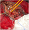
Anatomical bisegmentectomy of VI–VII in patient with metastases from kidney cancer.
Figure 2
Figure 2
Anatomical segmentectomy of VI in colorectal liver metastasis (white arrow). Portal vein branch of VI–VII (red arrow).
Figure 3
Figure 3
Multiple parenchymal sparing resections in patient with metastases from colorectal cancer. 1: Atypical resection of seg V; 2: Atypical resection of seg V-VI and 3: Atypical resection of segment VII.
Figure 4
Figure 4
US (A) assessment and CT (BD) assessment of Figure 3, patient. On portal phase of contrast medium evaluation, arrows ((B): V seg; (C): VII seg and (D): VI seg) show typical fluid collection due to surgical procedure.
Figure 5
Figure 5
In (A), atypical liver resection of lesion located in V-VIII segment in patient with metastases from colorectal cancer. In (B,C) CT post treatment evaluation. At discharge, arrow shows fluid collection; at 3 month CT follow-up, arrow shows scar tissue (C).
Figure 6
Figure 6
Active bleeding. During arterial CT assessment (A) and portal evaluation (B), arrow shows active overflow of contrast material.
Figure 7
Figure 7
Post-surgical MRI assessment. In (A) T2-W sequences shows (arrow) fluid collection. In (B), On EOB-phase, bile leak is detected as an active overflow of contrast material outside biliary tree and inside the fluid collection (arrow). In (C) EOB-phase post bile leak treatment evaluation, no overflow of contrast medium.
Figure 8
Figure 8
In (A,B) robotic atypical resection of lesion located in segment VIII. In (CE), CT evaluation: arrows show fluid collection.
Figure 9
Figure 9
Robotic Anatomycal S2 segmentectomy in patient with HCC (A). In (B,C), CT assessment at 3 months follow-up: arrows show scar tissue.

References

    1. Lafaro K.J., Stewart C., Fong A., Fong Y. Robotic Liver Resection. Surg. Clin. N. Am. 2020;100:265–281. doi: 10.1016/j.suc.2019.11.003. - DOI - PubMed
    1. Agarwal V., Divatia J.V. Enhanced recovery after surgery in liver resection: Current concepts and controversies. Korean J. Anesthesiol. 2019;72:119–129. doi: 10.4097/kja.d.19.00010. - DOI - PMC - PubMed
    1. Izzo F., Granata V., Grassi R., Fusco R., Palaia R., Delrio P., Carrafiello G., Azoulay D., Petrillo A., Curley S.A. Radiof-requency Ablation and Microwave Ablation in Liver Tumors: An Update. Oncologist. 2019;24:e990–e1005. doi: 10.1634/theoncologist.2018-0337. - DOI - PMC - PubMed
    1. Granata V., Grassi R., Fusco R., Setola S.V., Belli A., Ottaiano A., Nasti G., La Porta M., Danti G., Cappabianca S., et al. Intrahepatic cholangiocarcinoma and its differential diagnosis at MRI: How radiologist should assess MR features. Radiol. Med. 2021;126:1584–1600. doi: 10.1007/s11547-021-01428-7. - DOI - PubMed
    1. Ruan S.M., Huang H., Cheng M.Q., Lin M.X., Hu H.T., Huang Y., Li M.D., Lu M.D., Wang W. Shear-wave elastography combined with contrast-enhanced ultrasound algorithm for noninvasive characterization of focal liver lesions. Radiol. Med. 2022 doi: 10.1007/s11547-022-01575-5. - DOI - PubMed

LinkOut - more resources