. 2023 Jan 4;9(1):74.
doi: 10.3390/jof9010074.

The ERAD Pathway Participates in Fungal Growth and Cellulase Secretion in Trichoderma reesei

Affiliations

The ERAD Pathway Participates in Fungal Growth and Cellulase Secretion in Trichoderma reesei

Cheng Yao et al. J Fungi (Basel). .

Abstract

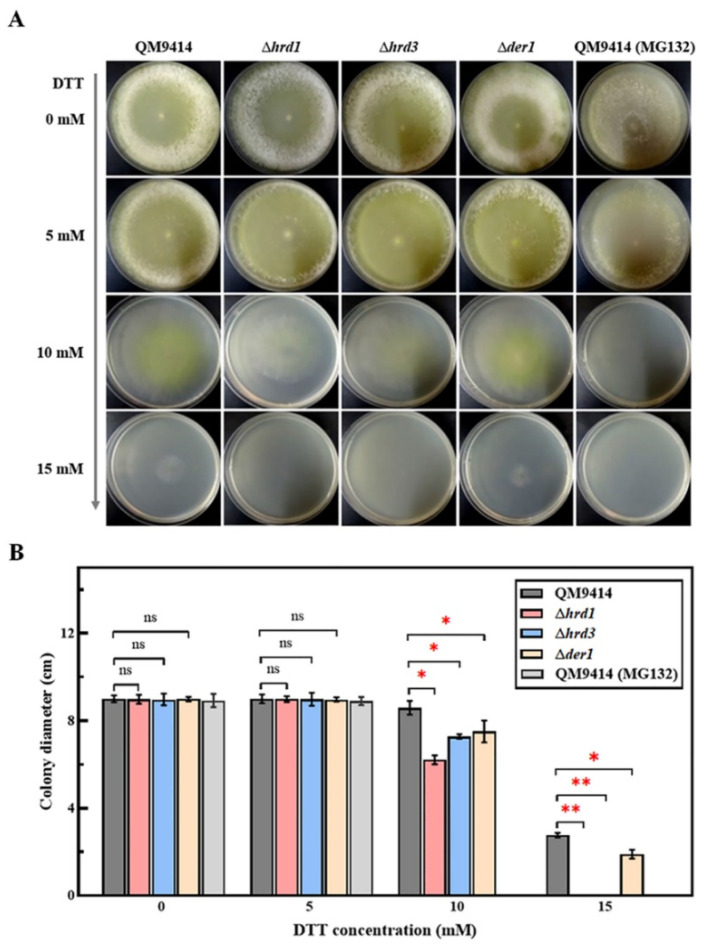
Trichoderma reesei is a powerful fungal cell factory for the production of cellulolytic enzymes due to its outstanding protein secretion capacity. Endoplasmic reticulum-associated degradation (ERAD) plays an integral role in protein secretion that responds to secretion pressure and removes misfolded proteins. However, the role of ERAD in fungal growth and endogenous protein secretion, particularly cellulase secretion, remains poorly understood in T. reesei. Here, we investigated the ability of T. reesei to grow under different stresses and to secrete cellulases by disrupting three major genes (hrd1, hrd3 and der1) involved in the critical parts of the ERAD pathway. Under the ER stress induced by high concentrations of DTT, knockout of hrd1, hrd3 and der1 resulted in severely impaired growth, and the mutants Δhrd1 and Δhrd3 exhibited high sensitivity to the cell wall-disturbing agents, CFW and CR. In addition, the absence of either hrd3 or der1 led to the decreased heat tolerance of this fungus. These mutants showed significant differences in the secretion of cellulases compared to the parental strain QM9414. During fermentation, the secretion of endoglucanase in the mutants was essentially consistent with that of the parental strain, while cellobiohydrolase and β-glucosidase were declined. It was further discovered that the transcription levels of the endoglucanase-encoding genes (eg1 and eg2) and the cellobiohydrolase-encoding gene (cbh1) were not remarkedly changed. However, the β-glucosidase-encoding gene (bgl1) was significantly downregulated in the ERAD-deficient mutants, which was presumably due to the activation of a proposed feedback mechanism, repression under secretion stress (RESS). Taken together, our results indicate that a defective ERAD pathway negatively affects fungal growth and cellulase secretion, which provides a novel insight into the cellulase secretion mechanism in T. reesei.

Keywords: ERAD; Trichoderma reesei; cellulase secretion; endoplasmic reticulum stress; fungal growth.


Conflict of interest statement

The authors declare that they have no competing interest.

Figures

Figure 1
Figure 1
Colony morphology and growth of the ERAD-deficient mutants under different concentrations of the stress agent DTT. (A) Plate photographs show the morphology of the control strains and the genetically manipulated ERAD-deficient mutants with increasing concentrations of DTT. The DTT concentrations added to the medium were 0, 5, 10 and 15 mM, respectively. The parental strain QM9414 treated with the proteasome inhibitor MG132 was used as the control. The strains were grown on MM containing glucose as the carbon source at 30 °C for 3 days. (B) Colony diameter of QM9414 (treated without/with MG132) and the ERAD-deficient mutants with increasing concentrations of DTT. Values represent the mean of three repeated measurements taken from at least three parallel experiments. The error bars refer to the standard deviations. The difference between the parental strain and the knockout strains was shown by analysis of ANOVA followed by Tukey’s test. * p < 0.05; ** p < 0.01. ns, not significant.
Figure 2
Figure 2
Cell wall stability of the ERAD-deficient mutants under ER pressure. Growth analysis of QM9414 and the ERAD-deficient mutants using the MM plates supplemented with different concentrations of Calcofluor white (A) or Congo red (B). The concentrations of CFW or CR were 0, 20, 40, and 80 μg/mL, respectively. All plates contained 10 mM DTT to induce ER pressure. The strains were grown on MM containing glucose as the carbon source at 30 °C for 3 days, and the diameters of all colonies were measured. Values represent the mean of three repeated measurements taken from at least three parallel experiments. The error bars refer to the standard deviations. The difference between the parental strain and the knockout strains was shown by analysis of ANOVA followed by Tukey’s test. * p < 0.05; ** p < 0.01. ns, not significant.
Figure 3
Figure 3
Thermal tolerance of the ERAD-deficient mutants at different culture temperatures. (A) The parental strain QM9414 and the ERAD-deficient mutants were grown on MM containing glucose as the carbon source and incubated under the given temperature conditions (25, 30, 37 °C). Growth was monitored for 3 days. (B) The colony diameters of QM9414 and the ERAD-deficient mutants were measured at different culture temperatures. Values represent the mean of three repeated measurements taken from at least three parallel experiments. The error bars refer to the standard deviations. The difference between the parental strain and the knockout strains was shown by analysis of ANOVA followed by Tukey’s test. ** p < 0.01. ns, not significant.
Figure 4
Figure 4
Cellulase secretion of the ERAD-deficient mutants under cellulase induction conditions. The endo-β-1,4-glucanase (EG) activity (A), the cellobiohydrolase (CBH) activity (B) and the β-glucosidase (BGL) activity (C) were detected in fermentation supernatants on the 3rd, 5th and 7th days. The parental strain QM9414 and the ERAD-deficient mutants were cultivated in a cellulase-inducing medium containing 2% microcrystalline cellulose and 2% corn steep liquor at 30 °C. (D) Intuitive analysis of the secreted β-glucosidase activity in the CMC-esculin plates experiment. The secretion of β-glucosidase was evaluated by measuring the diameter of the resulting black zone. Values represent the mean of three repeated measurements taken from at least three parallel experiments. The error bars refer to the standard deviations. The difference between the parental strain and the knockout strains was shown by analysis of ANOVA followed by Tukey’s test. * p < 0.05; ** p < 0.01. ns, not significant.
Figure 5
Figure 5
The ER pressure of the ERAD-deficient mutants under cellulase-induced secretion conditions. (A) The relative transcription levels of UPR-related genes bip1 and pdi1 in the ERAD-deficient mutants under cellulase-induced secretion conditions. (B) The relative transcription levels of hrd1/hrd3/der1 in the ERAD-deficient mutants under cellulase-induced secretion conditions. Cellulase induction medium contained 2% microcrystalline cellulose and 2% corn steep liquor, which promotes the production of large amounts of cellulase. The relative transcription level of each gene was calculated by the qPCR results of the fermentation samples on the 3rd, 5th, and 7th days. Actin gene was universally used to normalize gene transcription levels. Values represent the mean of three repeated measurements taken from at least three parallel experiments. The error bars refer to the standard deviations. The difference between the parental strain and the knockout strains was shown by analysis of ANOVA followed by Tukey’s test. * p < 0.05; ** p < 0.01. ns, not significant.
Figure 6
Figure 6
The transcription levels of cellulase component genes in ERAD-deficient mutants under cellulase-induced secretion conditions. (A) The relative transcription level of endoglucanase I gene eg1 in the ERAD-deficient mutants under cellulase-induced secretion conditions. (B) The relative transcription level of endoglucanase II gene eg2 in the ERAD-deficient mutants under cellulase-induced secretion conditions. (C) The relative transcription level of cellobiohydrolase I gene cbh1 in the ERAD-deficient mutants under cellulase-induced secretion conditions. (D) The relative transcription level of β-glucosidase gene bgl1 in the ERAD-deficient mutants under cellulase-induced secretion conditions. Cellulase induction medium contained 2% microcrystalline cellulose and 2% corn steep liquor, which promoted the production of large amounts of cellulase. The relative transcription level of each gene was calculated by the qPCR results of the fermentation samples on the 3rd, 5th, and 7th days. Actin gene was universally used to normalize gene transcription levels. Values represent the mean of three repeated measurements taken from at least three parallel experiments. The error bars refer to the standard deviations. The difference between the parental strain and the knockout strains was shown by analysis of ANOVA followed by Tukey’s test. * p < 0.05. ns, not significant.
Figure 7
Figure 7
The mechanism diagram of cellulase secretion changes caused by the deletion of critical single ERAD components under high cellulase secretion conditions in T. reesei. When the newly synthesized cellulase is translocated across the ER membrane co-translationally via the ribosome-sec61 translocation machinery, it undergoes a series of protein folding and post-translational modifications, such as molecular chaperone binding, disulfide bond formation and glycosylation, etc. Properly folded cellulase exits the ER, while unfolded or misfolded cellulase is recognized by Yos9 and directed to the ERAD pathway for degradation, where it is dislocated from the ER mediated by the Hrd1 ubiquitin ligase complex (Hrd1, Hrd3, Der1) and degraded by the cytosolic ubiquitin–proteasome system. Furthermore, when the baroreceptor Ire1 on the ER membrane senses a large number of unfolded and misfolded proteins, the UPR pathway, including Bip dissociation, Ire1 dimer phosphorylation, Hac1 translation and binding to the UPRE region, is initiated and upregulates molecular chaperones and folding enzymes expression, such as Bip1 and Pdi1. Under the ER pressure caused by the single-component deletion of the Hrd1 complex (Hrd1/Hrd3/Der1), unfolded and misfolded cellulases accumulate, which strongly activates the UPR, ERAD and RESS pathways. ERAD single-component deletion strengthens other components to make up for its absence, possibly resulting in degradation of cellobiohydrolase and decreased secretion. At this time, the ER stress prompts the cell to activate the RESS pathway, which may significantly reduce the expression and secretion of β-glucosidase. Gray cross means that the ERAD component (Hrd1/Hrd3/Der1) has been knocked out. Red dotted line represents the impact caused by ERAD single-component deletion. Red up and down arrows respectively indicate the increase and decrease in elements after ERAD single-component deletion.

Similar articles

Cited by

References

    1. Liu G., Qu Y. Integrated engineering of enzymes and microorganisms for improving the efficiency of industrial lignocellulose deconstruction. Eng. Microbiol. 2021;1:100005. doi: 10.1016/j.engmic.2021.100005. - DOI
    1. Garvey M., Klose H., Fischer R., Lambertz C., Commandeur U. Cellulases for biomass degradation: Comparing recombinant cellulase expression platforms. Trends Biotechnol. 2013;31:581–593. doi: 10.1016/j.tibtech.2013.06.006. - DOI - PubMed
    1. Filiatrault-Chastel C., Heiss-Blanquet S., Margeot A., Berrin J.G. From fungal secretomes to enzymes cocktails: The path forward to bioeconomy. Biotechnol. Adv. 2021;52:107833. doi: 10.1016/j.biotechadv.2021.107833. - DOI - PubMed
    1. Xue X., Wu Y., Qin X., Ma R., Luo H., Su X., Yao B. Revisiting overexpression of a heterologous β-glucosidase in Trichoderma reesei: Fusion expression of the Neosartorya fischeri Bgl3A to cbh1 enhances the overall as well as individual cellulase activities. Microb. Cell Fact. 2016;15:122. doi: 10.1186/s12934-016-0520-9. - DOI - PMC - PubMed
    1. Kubicek C.P., Mikus M., Schuster A., Schmoll M., Seiboth B. Metabolic engineering strategies for the improvement of cellulase production by Hypocrea jecorina. Biotechnol. Biofuels. 2009;2:19. doi: 10.1186/1754-6834-2-19. - DOI - PMC - PubMed

LinkOut - more resources