. 2023 Jan 10;13(1):202.
doi: 10.3390/life13010202.

Widefield and Ultra-Widefield Retinal Imaging: A Geometrical Analysis

Affiliations

Widefield and Ultra-Widefield Retinal Imaging: A Geometrical Analysis

Amedeo Lucente et al. Life (Basel). .

Abstract

Diabetic retinopathy (DR) often causes a wide range of lesions in the peripheral retina, which can be undetected when using a traditional fundus camera. Widefield (WF) and Ultra-Widefield (UWF) technologies aim to significantly expand the photographable retinal field. We conducted a geometrical analysis to assess the field of view (FOV) of WF and UWF imaging, comparing it to the angular extension of the retina. For this task, we shot WF images using the Zeiss Clarus 500 fundus camera (Carl Zeiss Meditec, Jena, Germany). Approximating the ocular bulb to an ideal sphere, the angular extension of the theoretically photographable retinal surface was 242 degrees. Performing one shot, centered on the macula, it was possible to photograph a retinal surface of ~570 mm2, with a FOV of 133 degrees. Performing four shots with automatic montage, we obtained a retinal surface area of ~1100 mm2 and an FOV of 200 degrees. Finally, performing six shots with semi-automatic montage, we obtained a retinal surface area of ~1400 mm2 and an FOV of 236.27 degrees, which is close to the entire surface of the retina. WF and UWF imaging allow the detailed visualization of the peripheral retina, with significant impact on the diagnosis and management of DR.

Keywords: diabetic retinopathy; field of view; imaging; retina; ultra-widefield; widefield.


Conflict of interest statement

The authors declare no conflict of interest.

Figures

Figure 1
Figure 1
Methods for assessing the angular field of view (FOV) in retinal imaging. (A) The FOV assessed by visual-angle (θv). The FOV angle is subtended by the imaged retinal region at the exit pupil of the eye, according to ISO 10940:2009 standards. (B) The FOV assessed by eye-angle (θe). The FOV angle is subtended by the imaged retinal region at the spherical center of the eye, that is, the insertion of the vertical diameter of the eyeball and the visual axis.
Figure 2
Figure 2
A single shot, centered on the macula. Photographable retinal linear length = 27.25 mm. FOV ≈ 133 × 133 degrees. Retinal surface ≈ 570 mm2.
Figure 3
Figure 3
Two shots, combined by automatic montage. Maximum photographable retinal linear length = 42.34 mm. FOV ≈ 200 × 133 degrees (wide by tall).
Figure 4
Figure 4
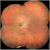
Four shots, combined by automatic montage. Maximum photographable retinal linear length = 41.39 mm. FOV ≈ 200 × 200 degrees (wide by tall). Retinal surface ≈ 1100 mm2.
Figure 5
Figure 5
Six shots, combined by semi-automatic montage. Maximum photographable retinal linear length = 49.46 mm. Maximum FOV ≈ 236.27 degrees. Retinal surface ≈ 1406.39 mm2.

Similar articles

Cited by

References

    1. Kumar V., Surve A., Kumawat D., Takkar B., Azad S., Chawla R., Shroff D., Arora A., Singh R., Venkatesh P. Ultra-wide field retinal imaging: A wider clinical perspective. Indian J. Ophthalmol. 2021;69:824–835. doi: 10.4103/ijo.IJO_1403_20. - DOI - PMC - PubMed
    1. Rivero M.E., Bartsch D.U., Otto T., Freeman W.R. Automated scanning laser ophthalmoscope image montages of retinal diseases. Ophthalmology. 1999;106:2296–2300. doi: 10.1016/S0161-6420(99)90529-8. - DOI - PubMed
    1. Early Treatment Diabetic Retinopathy Study Research Group Early Treatment Diabetic Retinopathy Study Design and Baseline Patient Characteristics: ETDRS Report Number 7. Ophthalmology. 1991;98:741–756. doi: 10.1016/S0161-6420(13)38009-9. - DOI - PubMed
    1. Kárason K.T., Vo D., Grauslund J., Rasmussen M.L. Comparison of different methods of retinal imaging for the screening of diabetic retinopathy: A systematic review. Acta Ophthalmol. 2022;100:127–135. doi: 10.1111/aos.14767. - DOI - PubMed
    1. Toslak D., Chau F., Erol M.K., Liu C., Chan R.V.P., Son T., Yao X. Trans-pars-planar illumination enables a 200° ultra-wide field pediatric fundus camera for easy examination of the retina. Biomed. Opt. Express. 2019;11:68–76. doi: 10.1364/BOE.11.000068. - DOI - PMC - PubMed

LinkOut - more resources