Review
. 2022 Dec 25;13(1):35.
doi: 10.3390/metabo13010035.

Genetic Predisposition to Hepatocellular Carcinoma

Affiliations
Review

Genetic Predisposition to Hepatocellular Carcinoma

Rosa M Pascale et al. Metabolites. .

Abstract

Liver preneoplastic and neoplastic lesions of the genetically susceptible F344 and resistant BN rats cluster, respectively, with human HCC with better (HCCB) and poorer prognosis (HCCP); therefore, they represent a valid model to study the molecular alterations determining the genetic predisposition to HCC and the response to therapy. The ubiquitin-mediated proteolysis of ERK-inhibitor DUSP1, which characterizes HCC progression, favors the unrestrained ERK activity. DUSP1 represents a valuable prognostic marker, and ERK, CKS1, or SKP2 are potential therapeutic targets for human HCC. In DN (dysplastic nodule) and HCC of F344 rats and human HCCP, DUSP1 downregulation and ERK1/2 overexpression sustain SKP2-CKS1 activity through FOXM1, the expression of which is associated with a susceptible phenotype. SAM-methyl-transferase reactions and SAM/SAH ratio are regulated by GNMT. In addition, GNMT binds to CYP1A, PARP1, and NFKB and PREX2 gene promoters. MYBL2 upregulation deregulates cell cycle and induces the progression of premalignant and malignant liver. During HCC progression, the MYBL2 transcription factor positively correlates with cells proliferation and microvessel density, while it is negatively correlated to apoptosis. Hierarchical supervised analysis, regarding 6132 genes common to human and rat liver, showed a gene expression pattern common to normal liver of both strains and BN nodules, and a second pattern is observed in F344 nodules and HCC of both strains. Comparative genetics studies showed that DNs of BN rats cluster with human HCCB, while F344 DNs and HCCs cluster with HCCP.

Keywords: BN rats; CYP1A; DUSP1; F344 rats; FOXM1; GNMT; MYBL2 transcription factor; PARP1 NFKB and PREX2 genes; hepatocellular carcinoma.


Conflict of interest statement

The authors declare no conflict of interest.

Figures

Figure 1
Figure 1
Metabolic cycles implicated in methionine metabolism, and long-range interactions of SAM. The inhibitory effects are indicated in red for SAM; and in green for SAH. The activator effects of SAM are shown in blue. Substrates: BET, betaine; CHOL, choline; DMG, dimethylglycine; dSAM, S-adenosylmethionine decarboxylated; GN, glycine; GSH, reduced glutathione; HCY, homocysteine; MTHF, methyltetrahydrofolate; MeTHF, 5,10-methenyl-tetrahydrofolate; PC, phosphatidylcholine; PE, phosphatidylethanolamine; SAH, S-adenosylhomocysteine; SAM S-adenosylmethionine; SN, sarcosine; THF, tetrahydrofolate. Enzymes: BAD, betaine aldehyde dehydrogenase; BHMT, betaine homocysteine-methyltransferase; CO, choline oxidase; DMGDH, dimethylglycine dehydrogenase; GNMT, glycine N-methyltransferase; MATI/III, MATII, methyladenosyltransferases I/III and II; MS, Methionine synthetase; MT, methyltransferases; MTHFR, methyltetrahydrofolate reductase; MeTHFR, 5,10-methenyl-tetrahydrofolate reductase; SAHH S-adenosylhomocysteine hydroxylase; SARDH, sarcosine dehydrogenase.
Figure 2
Figure 2
Effects of FOXM1: activation of CKS1 and SKP2 expression and CKS1/SKP2 complex formation, also known as “Ubiquitin ligase”, with consequent phosphorylated DUSP1 proteasomal degradation, and activation of Cdc2/25B/Cyclin B1, NEK2, and EPO.
Figure 3
Figure 3
Cell cycle protection from inhibition by P16INK4A through the complex CDC37/HSP90 and CRM1. The complex P16/E4 by sequestering CDK4/6, impedes the formation of the complex CDK4/6-CyclinD2 and consequently inhibits cell cycle.
Figure 4
Figure 4
Effect of HIF-1a on hepatocarcinogenesis. The HIF-1a axis sustains, together with TNF-a, FOXM1 expression and tumorigenicity. HIF-1a binding to MAT2A promoter inhibits SAM production and the consequent ERK1/2 inhibition by SAM, while it favors epithelial/mesenchymal transition (EMT) and tumorigenesis.
Figure 5
Figure 5
Effects of GNMT on CYP1A1, PARP-1, NFKB, and PRERX2, and its contribution to tumor prevention, gluconeogenesis, lipid, and glucose homeostasis.
Figure 6
Figure 6
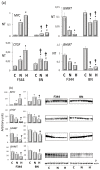
(a): qPCR analysis of Myc, Ctgf, Gnmt, and Bhmt RNA expression in normal liver (C), dysplastic nodules (N), and HCC (H) of F344 and BN rats. Results are means ± SD of six normal livers, 15 DNs 32 weeks after initiation, and 14 HCCs per strain. Number Target (NT). NT = 2-ΔCt, ΔCt = CT (target)–CT(RNR-18). Tukey–Kramer test: N and H vs. C, BN vs. F344, at least p < 0.05 for all genes tested. (b): Western blot of c-Myc, Ctgf, Gnmt, and Bhmt proteins expression. Optical densities of the peaks were normalized to β-actin values and expressed as arbitrary units. Data are means ± SD of three normal livers, five DNs, and five moderately-differentiated HCCs from F344 and BN rats. Tukey–Kramer test: (*) N and H vs. C, at least p < 0.05. (†) F344 vs. BN, p < 0.001.
Figure 7
Figure 7
Structure of MYB family members and interacting proteins. MYB family members are shown: they include MYBL1 and MYBL2. MYB-binding domain, made up of the three repeats R1, R2, and R3 binding to several proteins, among which include p100, PARP, Cyp40, c-Ski, N-CoR, C/EMPbeta, RAR, SMRT, and mSin3A. The MYBL1 and MBL2 protein structures are also shown. MYB co-activators are listed in green. MYB co-repressors are also listed.
Figure 8
Figure 8
Schematic representation of multistage hepatocarcinogenesis. Most initiated cells undergo death by apoptosis or re-differentiate to normal appearing phenotype, depending on the gravity of the carcinogenic stimulus. A strong stimulus irreversibly affects DNA resulting in cell death, whereas a weak stimulus can allow DNA repair and redifferentiation (retroverted arrows). Intermediate stimuli could be compatible with cell survival, and possible cell transformation. On the other hand, moderate DNA damage may induce carcinogenesis initiation. Several rounds of cell division, during the selective expansion of initiated cells, trigger the development of foci of altered hepatocytes (FAH), early dysplastic nodules (DNs), late DNs, and hepatocellular carcinomas (HCC).
Figure 9
Figure 9
Comparative functional genomics approach by integrated unsupervised hierarchical cluster analysis of 28 human surrounding non-tumorous livers, 35 HCCB, 35 HCCP, and rat surrounding liver, DN and HCC. Abbreviations: hSL and rSL, human and rat surrounding liver, hHCCB, human HCC with better prognosis; rBN DN, BN rat dysplastic nodules; rF344 DN, F344 rats dysplastic nodules; rBN HCC, BN rats hepatocellular carcinomas; hHCCP, human hepatocellular carcinomas with poorer prognosis; rF344 HCC, F344 rats hepatocellular carcinomas.

References

    1. Llovet J.M., Zucman-Rossi J., Pikarsky E., Sangro B., Schwartz M., Sherman M., Gores G. Hepatocellular carcinoma. Nat. Rev. Dis. Primers. 2016;2:16018. doi: 10.1038/nrdp.2016.18. - DOI - PubMed
    1. Nault J.C., Ningarhari M., Rebouissou S., Zucman-Rossi J. The role of telomeres and telomerase in cirrhosis and liver cancer. Nat. Rev. Gastroenterol. Hepatol. 2019;16:544–558. doi: 10.1038/s41575-019-0165-3. - DOI - PubMed
    1. Zucman-Rossi J., Villanueva A., Nault J.C., Llovet J.M. Genetic Landscape and Biomarkers of Hepatocellular Carcinoma. Gastroenterology. 2015;149:1226–1239.e4. doi: 10.1053/j.gastro.2015.05.061. - DOI - PubMed
    1. Wang H., Chen L., Zhou T., Zhang Z., Zeng C. p53 Mutation at Serine 249 and Its Gain of Function Are Highly Related to Hepatocellular Carcinoma after Smoking Exposure. Public Health Genomics. 2021;24:171–181. doi: 10.1159/000516598. - DOI - PubMed
    1. Singh G., Yoshida E.M., Rathi S., Marquez V., Kim P., Erb S.R., Salh B.S. Biomarkers for hepatocellular cancer. World J. Hepatol. 2020;12:558–573. doi: 10.4254/wjh.v12.i9.558. - DOI - PMC - PubMed

LinkOut - more resources