Review
. 2022 Dec 30;13(1):60.
doi: 10.3390/metabo13010060.

Obstructive Sleep Apnea, Circadian Clock Disruption, and Metabolic Consequences

Affiliations
Review

Obstructive Sleep Apnea, Circadian Clock Disruption, and Metabolic Consequences

Mikołaj Malicki et al. Metabolites. .

Abstract

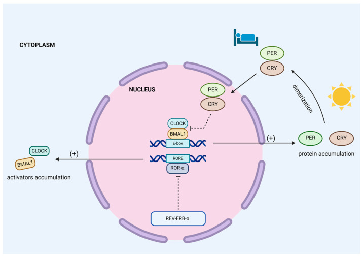
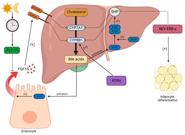
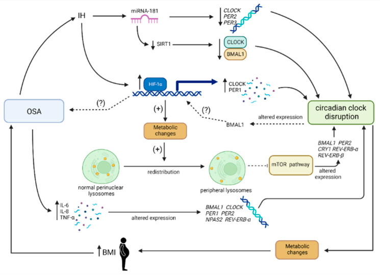
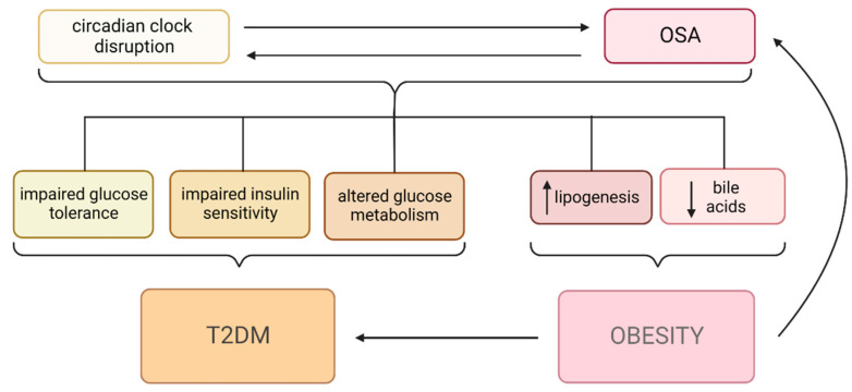
Obstructive sleep apnea (OSA) is a chronic disorder characterized by recurrent episodes of apnea and hypopnea during sleep. It is associated with various cardiovascular and metabolic complications, including type 2 diabetes mellitus (T2DM) and obesity. Many pathways can be responsible for T2DM development in OSA patients, e.g., those related to HIF-1 and SIRT1 expression. Moreover, epigenetic mechanisms, such as miRNA181a or miRNA199, are postulated to play a pivotal role in this link. It has been proven that OSA increases the occurrence of circadian clock disruption, which is also a risk factor for metabolic disease development. Circadian clock disruption impairs the metabolism of glucose, lipids, and the secretion of bile acids. Therefore, OSA-induced circadian clock disruption may be a potential, complex, underlying pathway involved in developing and exacerbating metabolic diseases among OSA patients. The current paper summarizes the available information pertaining to the relationship between OSA and circadian clock disruption in the context of potential mechanisms leading to metabolic disorders.

Keywords: OSA; circadian disruption; diabetes mellitus; metabolic complications; microRNA; obesity.


Conflict of interest statement

Authors declare no conflict of interest.

Figures

Figure 1
Figure 1
Core circadian clock mechanism. There are two main feedback loops. CLOCK and BMAL1 are transcription factors which act as activators. They work as heterodimers and bind to E-box sequences in DNA, leading to a large number of gene transcriptions, such as PER, CRY, and REV-ERB-α. PER and CRY proteins accumulate during the daily cycle in cells’ cytoplasm. Their highest levels occur before sleep. Following this process, they form a dimerm which is translocated to the nucleus. PER-CRY dimers exhibit repressor activity via the inhibition of CLOCK-BMAL1. This is a basic negative feedback loop that regulates the circadian clock. The second feedback mechanism of the core clock is composed of ROR-α and REV-ERB-α proteins. ROR-α binds to RORE sequences in DNA and promotes the transcription of activators such as CLOCK and BMAL1. REV-ERB-α is a repressor because it inhibits ROR-α activity. BMAL1—brain and muscle ARNT-like 1, CLOCK—clock circadian regulator/circadian locomotor output cycles protein kaput, CRY—cryptochrome, E-box—enhancer box, PER—period protein, REV-ERB-α—nuclear receptor subfamily 1 group D member 1, RORE—ROR response elements, ROR-α—nuclear retinoid-related orphan receptors α.
Figure 2
Figure 2
Impact of the circadian clock on lipid and bile acid homeostasis. Cholesterol is converted into bile acids in the biochemical pathway in hepatocytes. The main enzymes of this pathway are cholesterol 7 alpha-hydroxylase (CYP7A1) and sterol 12-alpha-hydroxylase (CYP8B1). CYP8B1 is stimulated by one of the circadian clock proteins, orphan receptor α (RORα). The production of bile acids is regulated by a negative feedback loop. Bile acids activate the nuclear farnesoid X receptor (FXR). It binds to the retinoid X receptor (RXR) and forms a heterodimer. The FXR-RXR complex activates a small heterodimer partner (SHP). The SHP protein inhibits CYP7A1 and CYP8B1 enzymes and decreases the production of bile acids. The circadian clock can influence the bile acid synthesis pathway via SHP. Specifically, REV-ERB-α inhibits SHP. Thus, REV-ERB-α promotes bile acid synthesis. Moreover, it stimulates adipocyte differentiation. Some bile acids are transported into the small intestine. They activate the FXR in enterocytes, resulting in the release of fibroblast growth factor 15 (FGF15). FGF15 promotes the fibroblast growth factor receptor-4/β-Klotho (FGFR4/β-Klotho) complex, inhibiting CYP7A1 and CYP8B1. This is the second negative feedback loop regulating bile acid synthesis. Day and night cycles can influence bile acid synthesis via the circadian regulation of Krupple-like factor 15 (KLF15) production. KLF15 restrains the release of FGF15 in enterocytes. CYP7A1—cholesterol 7 alpha-hydroxylase, CYP8B1—sterol 12-alpha-hydroxylase, FGF15—fibroblast growth factor 15, FGFR4—fibroblast growth factor receptor-4, FXR—nuclear farnesoid X receptor, KLF15—Krupple-like factor 15, REV-ERB-α—nuclear receptor subfamily 1 group D member 1, ROR-α—nuclear retinoid-related orphan receptors α, RXR—retinoid X receptor, SHP—small heterodimer partner, β-Klotho—β-Klotho.
Figure 3
Figure 3
The bidirectional link between OSA and circadian clock disruption. Obstructive sleep apnea (OSA) is associated with the occurrence of recurring intermittent hypoxia (IH) episodes. In such conditions, a miRNA-181 family, which is oxygen-sensitive, is overexpressed. This leads to the silencing of miRNA-181-targeted genes, including, among others, CLOCK, PER2, and PER3. Impaired clock gene fluctuations result in circadian clock disruption. Furthermore, miRNA-181 also targets the SIRT1 gene. SIRT1 deficiency leads to decreased CLOCK-BMAL1 levels, which triggers impaired circadian gene transcriptions and, finally, the development of circadian clock disruption. In oxygen-limited conditions, hypoxia-inducible factor-1α (HIF-1α) is upregulated. This results in increased levels of CLOCK and PER1 proteins. As mentioned above, changes in circadian clock gene products fluctuations leads to circadian clock disruptions. The overexpression of HIF-1α also generates metabolic changes. Subsequently, the altered spatial lysosome distribution in cells inhibits the mechanistic target of the rapamycin kinase (mTOR) pathway. It also induces changes in circadian clock gene levels. The HIF-1α-related pathway is most likely bidirectional. Circadian misalignment results in altered BMAL1 expression. This triggers HIF-1α upregulation, which probably promotes OSA development. Another pathway is associated with low-grade inflammation. OSA stimulates the overproduction of interleukin (IL) 6 and 8, as well as tumor necrosis factor-α (TNF-α). The basic opposite pathway is associated with metabolic changes resulting from a disordered circadian clock. This promotes an increase in body mass index (BMI), an established risk factor for OSA development. BMAL1—brain and muscle ARNT-like 1, BMI—body mass index, CLOCK—clock circadian regulator/circadian locomotor output cycles protein kaput, CRY1—cryptochrome 1, HIF-1α—hypoxia-inducible factor-1α, IH—intermittent hypoxia, IL-6—interleukin 6, IL-8—interleukin 8, NPAS2—neuronal PAS domain protein 2, miRNA-181—micro RNA-181, OSA—obstructive sleep apnea, PER1—period protein 1, PER2—period protein 2, PER3—period protein 3, REV-ERB-α—nuclear receptor subfamily 1 group D member 1, SIRT1—sirtuin 1, TNF-α—tumor necrosis factor-α.
Figure 4
Figure 4
Summary of relationships between obstructive sleep apnea (OSA), circadian clock disruption, and its clinical effects. The relationship between circadian clock disruption and OSA is bidirectional. They both lead to variable metabolic changes—impaired glucose tolerance, impaired insulin sensitivity, and altered glucose metabolism—which result in the development of diabetes mellitus type 2 (T2DM). Increased lipogenesis and decreased bile acid levels promote the development of obesity. Obesity is also an independent risk factor for both OSA and T2DM. OSA—obstructive sleep apnea, T2DM—diabetes mellitus type 2.

Similar articles

Cited by

References

    1. Rundo J.V. Obstructive Sleep Apnea Basics. Cleve. Clin. J. Med. 2019;86:2–9. doi: 10.3949/ccjm.86.s1.02. - DOI - PubMed
    1. Patel S.R. Obstructive Sleep Apnea. Ann. Intern. Med. 2019;171:ITC81–ITC96. doi: 10.7326/AITC201912030. - DOI - PubMed
    1. Semelka M., Wilson J., Floyd R. Diagnosis and Treatment of Obstructive Sleep Apnea in Adults. Am. Fam. Physician. 2016;94:355–360. doi: 10.6061/clinics/2013(08)17. - DOI - PubMed
    1. Young T., Evans L., Finn L., Palta M. Estimation of the Clinically Diagnosed Proportion of Sleep Apnea Syndrome in Middle-Aged Men and Women. Sleep. 1997;20:705–706. doi: 10.1093/sleep/20.9.705. - DOI - PubMed
    1. Punjabi N.M. The Epidemiology of Adult Obstructive Sleep Apnea. Proc. Am. Thorac. Soc. 2008;5:136–143. doi: 10.1513/pats.200709-155MG. - DOI - PMC - PubMed

LinkOut - more resources