

Development of Tailored Graphene Nanoparticles: Preparation, Sorting and Structure Assessment by Complementary Techniques
- PMID: 36677624
- PMCID: PMC9865347
- DOI: 10.3390/molecules28020565
Development of Tailored Graphene Nanoparticles: Preparation, Sorting and Structure Assessment by Complementary Techniques
Abstract
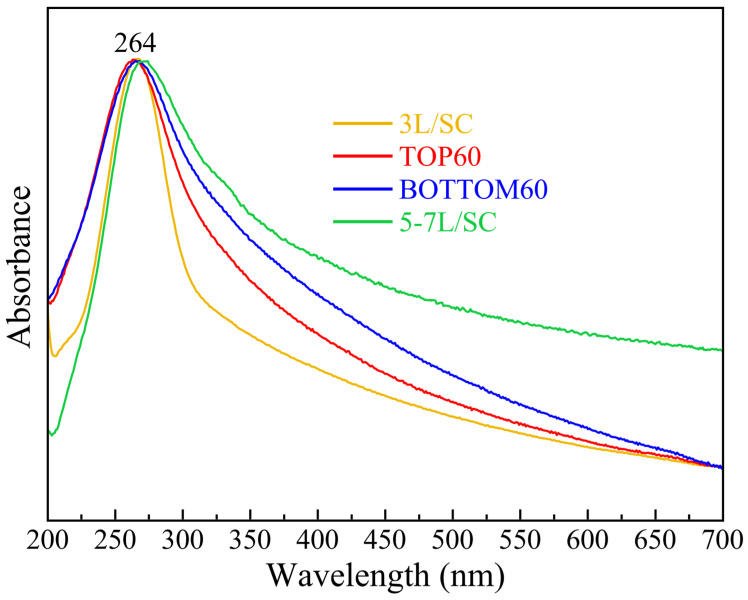
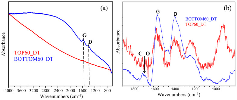
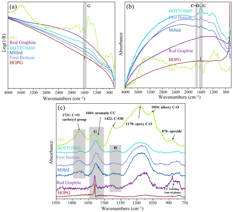
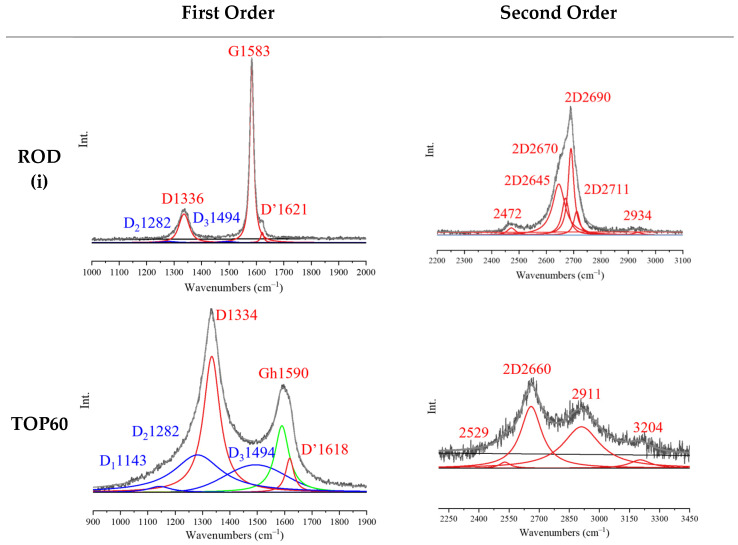
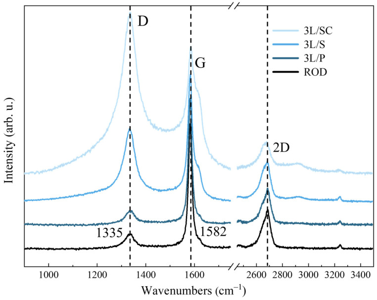
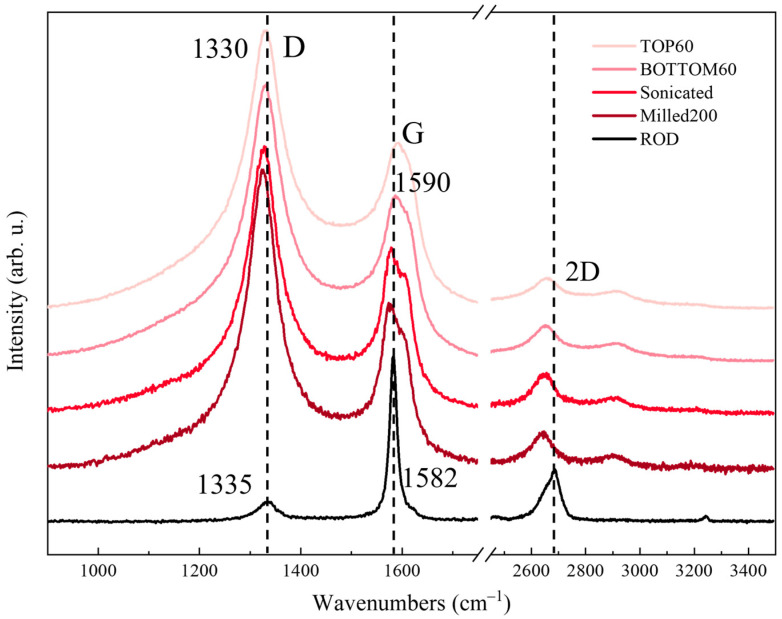
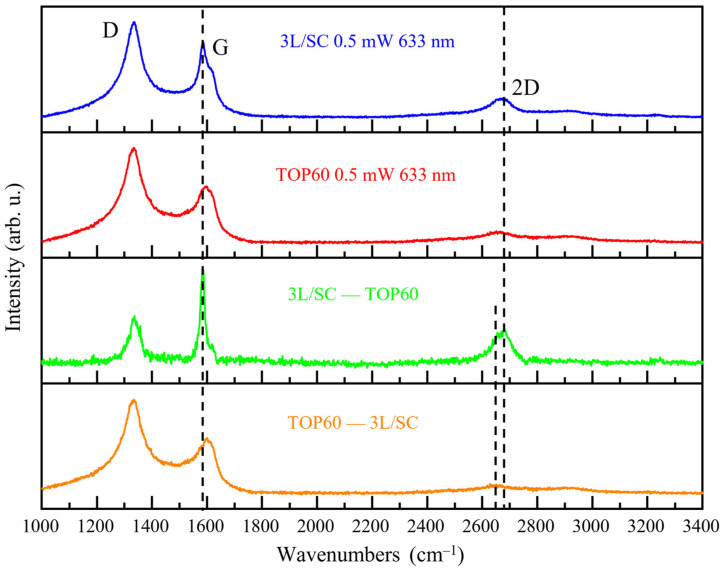
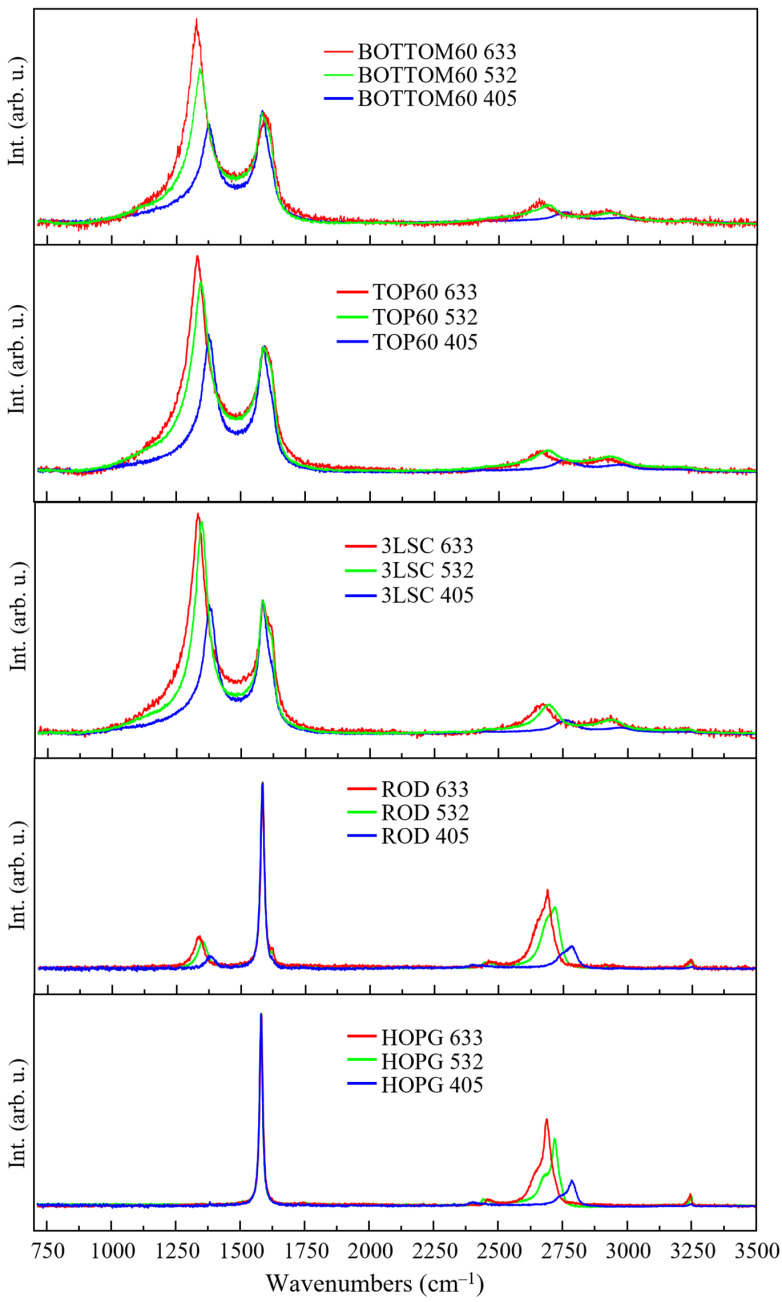
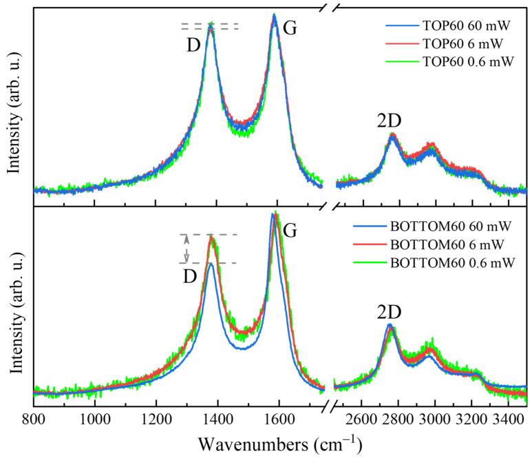
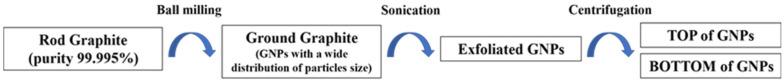
We present a thorough structural characterization of Graphene Nano Particles (GNPs) prepared by means of physical procedures, i.e., ball milling and ultra-sonication of high-purity synthetic graphite. UV-vis absorption/extinction spectroscopy, Dynamic Light Scattering, Transmission Electron Microscopy, IR and Raman spectroscopies were performed. Particles with small size were obtained, with an average lateral size <L> = 70−120 nm, formed by few <N> = 1−10 stacked layers, and with a small number of carboxylic groups on the edges. GNPs relatively more functionalized were separated by centrifugation, which formed stable water dispersions without the need for any surfactant. A critical reading and unified interpretation of a wide set of spectroscopic data was provided, which demonstrated the potential of Specular Reflectance Infrared Spectroscopy for the diagnosis and quantification of chemical functionalization of GNPs. Raman parameters commonly adopted for the characterization of graphitic materials do not always follow a monotonic trend, e.g., with the particle size and shape, thus unveiling some limitations of the available spectroscopic metrics. This issue was overcome thanks to a comparative spectra analysis, including spectra deconvolution by means of curve fitting procedures, experiments on reference materials and the exploitation of complementary characterization techniques.
Keywords: Raman spectroscopy; chemical functionalization; drug delivery platform; graphene-nanoparticles; infrared spectroscopy; transmission electron microscopy.
Conflict of interest statement
The authors declare no conflict of interest.
Figures
Similar articles
-
Tailored graphene nanoparticles for biomedical application: preliminary in vitro characterization of the functionality in model cell lines.Int J Pharm. 2024 Dec 25;667(Pt B):124914. doi: 10.1016/j.ijpharm.2024.124914. Epub 2024 Nov 7. Int J Pharm. 2024. PMID: 39515671
-
Studying disorder in graphite-based systems by Raman spectroscopy.Phys Chem Chem Phys. 2007 Mar 21;9(11):1276-91. doi: 10.1039/b613962k. Epub 2007 Jan 11. Phys Chem Chem Phys. 2007. PMID: 17347700 Review.
-
A facile and novel synthetic method for the preparation of hydroxyl capped fluorescent carbon nanoparticles.Colloids Surf B Biointerfaces. 2013 Feb 1;102:63-9. doi: 10.1016/j.colsurfb.2012.08.016. Epub 2012 Sep 1. Colloids Surf B Biointerfaces. 2013. PMID: 23006553
-
Physicochemical characteristics of pristine and functionalized graphene.J Appl Toxicol. 2017 Nov;37(11):1288-1296. doi: 10.1002/jat.3493. Epub 2017 Jul 5. J Appl Toxicol. 2017. PMID: 28677847
-
Raman scattering of non-planar graphite: arched edges, polyhedral crystals, whiskers and cones.Philos Trans A Math Phys Eng Sci. 2004 Nov 15;362(1824):2289-310. doi: 10.1098/rsta.2004.1442. Philos Trans A Math Phys Eng Sci. 2004. PMID: 15482980 Review.
Cited by
-
Insights into the Safety and Versatility of 4D Printed Intravesical Drug Delivery Systems.Pharmaceutics. 2023 Feb 24;15(3):757. doi: 10.3390/pharmaceutics15030757. Pharmaceutics. 2023. PMID: 36986618 Free PMC article.
References
-
- Potts J.R., Dreyer D.R., Bielawski C.W., Ruoff R.S. Graphene-based polymer nanocomposites. Polymer. 2011;52:5–25. doi: 10.1016/j.polymer.2010.11.042. - DOI
MeSH terms
Substances
Grants and funding
LinkOut - more resources
Full Text Sources
