. 2023 Jan 6;28(2):590.
doi: 10.3390/molecules28020590.

Synthesis, Biological Evaluation and Molecular Modeling Studies of Naphthoquinone Sulfonamides and Sulfonate Ester Derivatives as P2X7 Inhibitors

Affiliations

Synthesis, Biological Evaluation and Molecular Modeling Studies of Naphthoquinone Sulfonamides and Sulfonate Ester Derivatives as P2X7 Inhibitors

Paulo Anastácio Furtado Pacheco et al. Molecules. .

Abstract

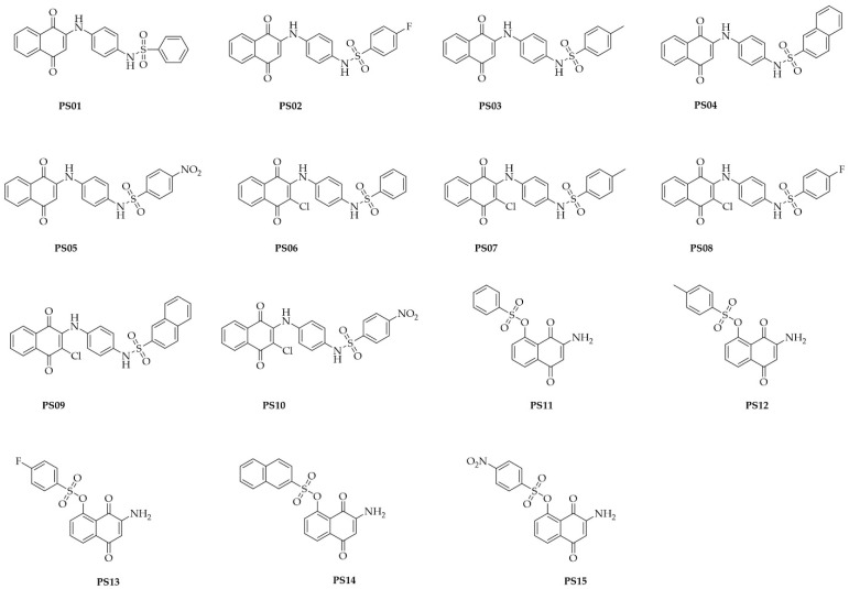
ATP acts in the extracellular environment as an important signal, activating a family of receptors called purinergic receptors. In recent years, interest in the potential therapeutics of purinergic components, including agonists and antagonists of receptors, has increased. Currently, many observations have indicated that ATP acts as an important mediator of inflammatory responses and, when found in high concentrations in the extracellular space, is related to the activation of the P2X7 purinergic receptor. In this sense, the search for new inhibitors for this receptor has attracted a great deal of attention in recent years. Sulfonamide derivatives have been reported to be potent inhibitors of P2X receptors. In this study, ten naphthoquinone sulfonamide derivatives and five naphthoquinone sulfonate ester derivatives were tested for their inhibitory activity on the P2X7 receptor expressed in peritoneal macrophages. Some compounds showed promising results, displaying IC50 values lower than that of A740003. Molecular docking and dynamic studies also indicated that the active compounds bind to an allosteric site on P2X7R. The binding free energy indicates that sulfonamides have an affinity for the P2X7 receptor similar to A740003. Therefore, the compounds studied herein present potential P2X7R inhibition.

Keywords: ATP; biomass; heterocycles; inflammation; naphthoquinones; sulfonamides.


Conflict of interest statement

The authors declare that they have no conflicts of interest.

Figures

Figure 1
Figure 1
Chemical structures of naphthoquinone sulfonamide and sulfonate ester derivatives tested in this study.
Figure 2
Figure 2
Quantification of cellular metabolic activity after treatment with the PS01–15 series employing the resazurin reduction capacity. Cells were treated with 10 µM of the substances and 0.5% TX−100 in the positive control (PC). Nontreated cells correspond to the negative control (NT). Comparison of NT vs. treatments. The error bar is representative of 3 experiments in triplicate on 3 different days. ANOVA (***) p < 0.0001.
Figure 3
Figure 3
P2X7 receptor−induced PI uptake. Peritoneal macrophages were pretreated for 10 min with sulfonamides at 10 µM and subsequently stimulated with 5 mM ATP for 15 min. As an inhibition control, the cells were treated with A740003 at 10 µM for 10 min. The positive control (PC) was 0.5% Triton−X 100. Nontreated cells correspond to the negative control (NT). The bar error is representative of 3 experiments in triplicate on 3 different days. ANOVA (***) p < 0.0001.
Figure 4
Figure 4
Dose−response curve for the sulfonamides. Peritoneal macrophages were pretreated for 10 min with sulfonamides at crescent concentrations and subsequently stimulated with 5 mM ATP for 15 min. As an inhibition control, the cells were treated with A740003 at 10 µM for 10 min (data not shown). The error bar is representative of 3 experiments in triplicate on 3 different days. M – drug concentration.
Figure 5
Figure 5
Dose-response curve for the sulfonamides. Peritoneal macrophages were pretreated for 24 h with sulfonamides at crescent concentrations. The error bar is representative of 3 experiments in triplicate on 3 different days. M—drug concentration.
Figure 5
Figure 5
Dose-response curve for the sulfonamides. Peritoneal macrophages were pretreated for 24 h with sulfonamides at crescent concentrations. The error bar is representative of 3 experiments in triplicate on 3 different days. M—drug concentration.
Figure 6
Figure 6
Anti−edematogenic effects of sulfonamides on ATP-induced paw edema in mice. Groups with five Swiss Webster mice treated with ATP (intra-plantar) were preincubated for 30 min with increasing doses of sulfonamides. All sulfonamide data were compared with crescent BBG doses (data not shown). Paw edema was measured at 30 min after ATP application. These results represent three distinct days with five animals for each group.
Figure 7
Figure 7
Superposition of the docked sulfonamide compounds into the P2X7 allosteric site. P2X7R is depicted in the cartoon, and each chain is represented in a different color. The sulfonamide ligands are depicted in gray, and the A740003 ligand is depicted in cyan. The main interacting residues are depicted in an enlarged view. The A740003 structure conformation was retrieved from PDB (ID: 5U1U).
Figure 8
Figure 8
RMSD (root-mean-square deviation) variation of the ligands (A740003, PS01, PS02, PS03, PS09, and PS10) in relation to the P2X7 receptor during 50 ns of molecular dynamics simulation.
Figure 9
Figure 9
Molecular docking of the sulfonamide derivatives into the P2X7 allosteric pocket. The P2X7R is depicted in cartoon and the main residue involved in the interaction is depicted in stick (A) In pink is represented the PS01 (B) In cyan is represented the PS02 (C) In dark blue is represented the PS03 (D) In brown is represented the PS09 (E) In yellow is represented the PS10.
Figure 10
Figure 10
Binding free energy fluctuation of the ligands (A740003, PS01, PS02, PS03, PS09, and PS10) into the P2X7 allosteric binding site during the last 5 ns of simulation for each complex trajectory. The blue line indicates the binding free energy fluctuation, and the dashed red line indicates the binding free energy average.

Similar articles

Cited by

References

    1. Burnstock G., Williams M. P2 purinergic receptors: Modulation of cell function and therapeutic potential. J. Pharmacol. Exp. Ther. 2000;295:862–869. - PubMed
    1. Ralevic V., Burnstock G. Receptors for purines and pyrimidines. Pharmacol. Rev. 1998;50:413–492. doi: 10.1007/978-3-642-28863-0_5. - DOI - PubMed
    1. Burnstock G. Purinergic signalling: From discovery to current developments. Exp. Physiol. 2014;99:16–34. doi: 10.1113/expphysiol.2013.071951. - DOI - PMC - PubMed
    1. Ijzerman A.P., Jacobson K.A., Müller C.E., Cronstein B.N., Cunha R.A. International Union of Basic and Clinical Pharmacology. CXII: Adenosine Receptors: A Further UpdateS. Pharmacol. Rev. 2022;74:340–372. doi: 10.1124/pharmrev.121.000445. - DOI - PMC - PubMed
    1. Illes P., Müller C.E., Jacobson K.A., Grutter T., Nicke A., Fountain S.J., Kennedy C., Schmalzing G., Jarvis M.F., Stojilkovic S.S., et al. Update of P2X receptor properties and their pharmacology: IUPHAR Review 30. Br. J. Pharmacol. 2020;178:489–514. doi: 10.1111/bph.15299. - DOI - PMC - PubMed

MeSH terms

Substances

LinkOut - more resources