. 2023 Jan 15;28(2):865.
doi: 10.3390/molecules28020865.

Development of [124/125I]IAZA as a New Proteinopathy Imaging Agent for Alzheimer's Disease

Affiliations

Development of [124/125I]IAZA as a New Proteinopathy Imaging Agent for Alzheimer's Disease

Thrisha T Reddy et al. Molecules. .

Abstract

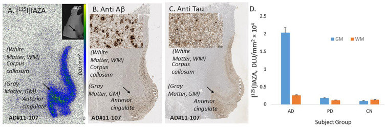
Radioiodinated imaging agents for Aβ amyloid plaque imaging in Alzheimer’s disease (AD) patients have not been actively pursued. Our previous studies employed the “diaza” derivatives [11C]TAZA and [18F]flotaza in order to develop successful positron emission tomography (PET) imaging agents for Aβ plaques. There is a need for radioiodinated imaging agents for Aβ plaques for single photon emission computed tomography (SPECT) and PET imaging. We report our findings on the preparation of [124/125I]IAZA, a “diaza” analog of [11C]TAZA and [18F]flotaza, and the evaluation of binding to Aβ plaques in the postmortem human AD brain. The binding affinity of IAZA for Aβ plaques was Ki = 10.9 nM with weak binding affinity for neurofibrillary tangles (Ki = 3.71 μM). Both [125I]IAZA and [124I]IAZA were produced in >25% radiochemical yield and >90% radiochemical purity. In vitro binding of [125I]IAZA and [124I]IAZA in postmortem human AD brains was higher in gray matter containing Aβ plaques compared to white matter (ratio of gray to white matter was >7). Anti-Aβ immunostaining strongly correlated with [124/125I]IAZA in postmortem AD human brains. The binding of [124/125I]IAZA in postmortem human AD brains was displaced by the known Aβ plaque imaging agents. Thus, radiolabeled [124/123I]IAZA may potentially be a useful PET or SPECT radioligand for Aβ plaques in brain imaging studies.

Keywords: Alzheimer’s disease; IAZA; imaging; iodine-124; iodine-125; postmortem human AD brain; transgenic 5xFAD mice; β-amyloid plaques.


Conflict of interest statement

The authors declare that the research was conducted in the absence of any commercial or financial relationships that could be construed as a potential conflict of interest.

Figures

Figure 1
Figure 1
Chemical structures of Aβ-plaque- and Tau-binding radioligands: 1. [124/125I]IBETA; 2. [125I]IPPI; 3. [11C]TAZA; 4. [18F]Flotaza; 5. [124/125I]IAZA.
Figure 2
Figure 2
In vitro competition of IAZA with [125I]IBETA and [125I]IPPI in postmortem human AD brain anterior cingulate (AC) and corpus callosum. (A) Brain slice of postmortem human AD 10 µm thick (inset shows scan anti-Aβ IHC labeled AC). (B) Adjacent slice of AC of same AD subject showing total binding of [125I]IBETA in Aβ-rich regions (arrows). (C) Same AD subject in the presence of 1 μM IAZA showing displacement of [125I]IBETA from AC. (D) Brain slice 10 µm thick of postmortem human AD subject (inset shows scan anti-Tau IHC labeled AC). (E) Adjacent slice of AC of same AD subject showing total binding of [125I]IPPI binding in Tau-rich regions (arrows); (F). Same AD subject in the presence of 10 μM IAZA showing partial displacement of [125I]IPPI from AC.
Figure 3
Figure 3
In vitro binding of [125I]IAZA in human postmortem AD, PD and CN brains consisting of anterior cingulate. (A) Postmortem human AD 10 µm thick brain slice shows [125I]IAZA binding mainly in the gray matter (GM) regions of anterior cingulate (AC) and none in the white matter (WM) regions of corpus callosum (CC). (B) Adjacent slice of the same subject immunostained with anti-Aβ showing abundance of Aβ plaques in the GM regions (inset shows magnified view of Aβ plaques). (C) Adjacent slice of the same subject immunostained with anti-Tau showing abundance of Tau in the GM regions (inset shows magnified view of Tau). (D) Plot shows high binding of [125I]IAZA in gray matter (GM) and white matter (WM) regions of AD subjects, while PD and CN subjects had little binding.
Figure 4
Figure 4
In vitro competition of [125I]IAZA with drugs in postmortem human AD brains containing temporal cortex and anterior cingulate showing binding in the gray matter (GM) and white matter (WM). All drugs used were at 10 μM concentration. Tau-binding drugs MK-6240 and IPPI reduced the binding to approximately 55%. Both MAO-A drug clorgyline and MAO-B drug deprenyl reduced the binding to approximately 65%. Drugs binding to Aβ plaques, IAZA and BrBETA, were able to displace most of the [125I]IAZA binding (>95%).
Figure 5
Figure 5
In vitro binding of [124I]IAZA in postmortem human AD brain. (A) Anti-Aβ IHC postmortem human AD 10 µm thick brain slice of temporal cortex (inset shows scan of brain slice). (B) Adjacent slice of temporal cortex of same AD subject showing [124I]IAZA binding in Aβ rich regions. (C) Human AD anterior cingulate (AC) and corpus callosum (CC) in 10 µm brain slice, inset shows anti-Aβ IHC in AC, where most Aβ plaques are located. (D) Adjacent slice of same AD subject showing [124I]IAZA binding in Aβ-rich regions in AC and none in CC. (E) A plot shows high [124I]IAZA binding in cortical regions of AD subjects, which was displaced by 10 μM of unlabeled IAZA.
Figure 6
Figure 6
[125I]NaI and [124I]NaI were used to prepare [125I]IAZA (8) and [124I]IAZA (9) by electrophilic substitution of the tributyltin derivative. TLC of [125I]IAZA shows the purity of >90%, and TLC of [124I]IAZA shows the purity of >90%.

References

    1. Braak H., Thal D.R., Ghebremedhin E., Del Tredici K. Stages of the pathologic process in Alzheimer disease: Age categories from 1 to 100 years. J. Neuropath. Exptl. Neurol. 2011;70:960–969. doi: 10.1097/NEN.0b013e318232a379. - DOI - PubMed
    1. Serrano-Pozo A., Frosch M.P., Masliah E., Hyman B.T. Neuropathological alterations in Alzheimer’s disease. Cold Spring Harb. Perspect. Med. 2011;1:a006189. doi: 10.1101/cshperspect.a006189. - DOI - PMC - PubMed
    1. Small D.H., McLean C.A. Alzheimer’s disease and the amyloid protein: What is the role of amyloid? J. Neurochem. 1999;73:443–449. doi: 10.1046/j.1471-4159.1999.0730443.x. - DOI - PubMed
    1. Oakley H., Cole S.L., Logan S., Maus E., Shao P., Craft J., Guillozet-Bongaarts A., Ohno M., Disterhoft J., Van Eldik L., et al. Intraneuronal β-amyloid aggregates, neurodegeneration, and neuron loss in transgenic mice with five familial Alzheimer’s disease mutations: Potential factors in amyloid plaque formation. J. Neurosci. 2006;26:10129–10140. doi: 10.1523/JNEUROSCI.1202-06.2006. - DOI - PMC - PubMed
    1. Hansson O., Lehmann S., Otto M., Zetterberg H., Lewczuk P. Advantages and disadvantages of the use of the CSF amyloid β (Aβ) 42/40 ratio in the diagnosis of Alzheimer’s disease. Alzheimer’s Res. Ther. 2019;11:1–15. doi: 10.1186/s13195-019-0485-0. - DOI - PMC - PubMed

MeSH terms