Review
. 2022 Dec 28;12(1):49.
doi: 10.3390/pathogens12010049.

Mouse Models for Mycobacterium tuberculosis Pathogenesis: Show and Do Not Tell

Affiliations
Review

Mouse Models for Mycobacterium tuberculosis Pathogenesis: Show and Do Not Tell

Pablo Soldevilla et al. Pathogens. .

Abstract

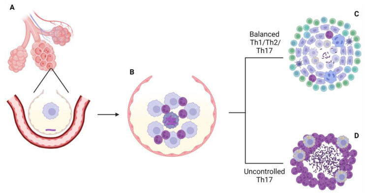
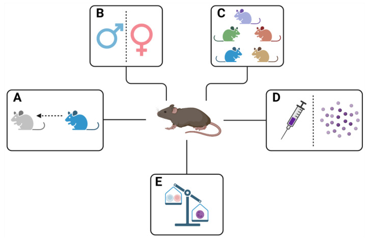
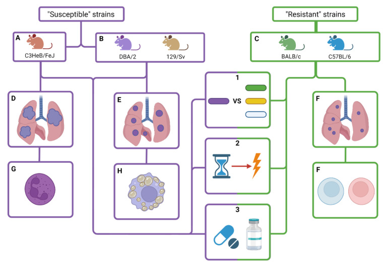
Science has been taking profit from animal models since the first translational experiments back in ancient Greece. From there, and across all history, several remarkable findings have been obtained using animal models. One of the most popular models, especially for research in infectious diseases, is the mouse. Regarding research in tuberculosis, the mouse has provided useful information about host and bacterial traits related to susceptibility to the infection. The effect of aging, sexual dimorphisms, the route of infection, genetic differences between mice lineages and unbalanced immunity scenarios upon Mycobacterium tuberculosis infection and tuberculosis development has helped, helps and will help biomedical researchers in the design of new tools for diagnosis, treatment and prevention of tuberculosis, despite various discrepancies and the lack of deep study in some areas of these traits.

Keywords: C3HeB/FeJ; chemotherapy; history; immunopathology; mouse; resistance; tolerance; tuberculosis; vaccines.


Conflict of interest statement

The authors declare no conflict of interest.

Figures

Figure 1
Figure 1
M. tuberculosis arrives to the host’s alveoli after inhalation of infected aerosols (A). There, the bacillus faces pulmonary surfactant secreted by type II pneumocytes and encounters the alveolar macrophage. Once it’s phagocytized, M. tuberculosis secretes ESAT-6 peptide to avoid the formation of phagolysosome and escape to the cytosol of the AM. That allows the bacillus to replicate and kill the AM. This causes the inflammation of the alveoli and the entrance of more AM and neutrophils (B). The inflammation leads to the drainage of alveolar fluid to the lymph nodes, where antigen presentation will occur. From there, two possible endings of the infection can happen: if there is a coordinated immune response between Th1, Th2 and Th17 responses, the infection will be controlled (C). If there is an excess of Th17 response, there will be excessive neutrophilia, disorganized granuloma formation and tissue destruction, causing the dissemination of the infection (D).
Figure 2
Figure 2
Representation of the traits that can influence development of tuberculosis: (A) Aging; (B) Differences between sexes; (C) Genetic differences between different lineages of mice; (D) The route of infection (intravenous or aerosol); (E) Immunocompromising elements that can unbalance the immune response against infections (e.g., diabetes, coinfections, etc.).
Figure 3
Figure 3
Although being tolerant hosts for M. tuberculosis infection, mice can be classified in two groups regarding tuberculosis development. There are “susceptible” mice, which are C3HeB/FeJ (A), DBA/2 and 129/Sv (B), and there are resistant mice, which are BALB/c and C57BL6 (C). C3HeB/FeJ mice are able to develop human-like active tuberculosis liquefied lesions (D) due to the hyper-inflammatory response upon the infection leaded, mainly, by neutrophilic infiltration (G); DBA/2 and 129/Sv mice are also classified as susceptible, but pulmonary lesions are not necrotic nor liquefied (E) and the cause of their susceptibility is the dissemination of infected foamy macrophages (H) to other lung areas. On the other hand, resistant mice BALB/c and C57BL/6 develop smaller lesions (F) than susceptible mice because the immune response in this case is leaded mainly by lymphocytes (F). Both susceptible and resistant types of mice can be used for: (1) Evaluating the virulence of different strains of M. tuberculosis and comparing them to more common lineages; (2) Studying the mechanisms that unleash the development of tuberculosis to the initial latent infection; (3) Testing new drugs, vaccines and treatments against M. tuberculosis.

References

    1. Debernardi A., Sala E., D’Aliberti G., Talamonti G., Franchini A.F., Collice M. Alcmaeon of Croton. Neurosurgery. 2010;66:247–252. doi: 10.1227/01.NEU.0000363193.24806.02. - DOI - PubMed
    1. Baumans V. Use of animals in experimental research: An ethical dilemma? Gene Ther. 2004;11:S64–S66. doi: 10.1038/sj.gt.3302371. - DOI - PubMed
    1. Gross C.G. Aristotle on the Brain. Neurosci. 1995;1:245–250. doi: 10.1177/107385849500100408. - DOI
    1. Bates A.W.H. Vivisection, Virtue, and the Law in the Nineteenth Century. Anti-Vivisect. Prof. Med. Br. 2017:13–41. doi: 10.1057/978-1-137-55697-4_2. - DOI
    1. la Follette H., Niall S. Animal experimentation: The legacy of Claude Bernard. Int. Stud. Philos. Sci. 1994;8:195–210. doi: 10.1080/02698599408573495. - DOI

LinkOut - more resources