. 2022 Dec 27;16(1):37.
doi: 10.3390/ph16010037.

Afatinib Reverses EMT via Inhibiting CD44-Stat3 Axis to Promote Radiosensitivity in Nasopharyngeal Carcinoma

Affiliations

Afatinib Reverses EMT via Inhibiting CD44-Stat3 Axis to Promote Radiosensitivity in Nasopharyngeal Carcinoma

Huichao Huang et al. Pharmaceuticals (Basel). .

Abstract

Background: Afatinib, a second-generation tyrosine kinase inhibitor (TKI), exerts its radiosensitive effects in nasopharyngeal carcinoma (NPC). However, the detailed mechanism of afatinib-mediated sensitivity to radiation is still obscure in NPC.

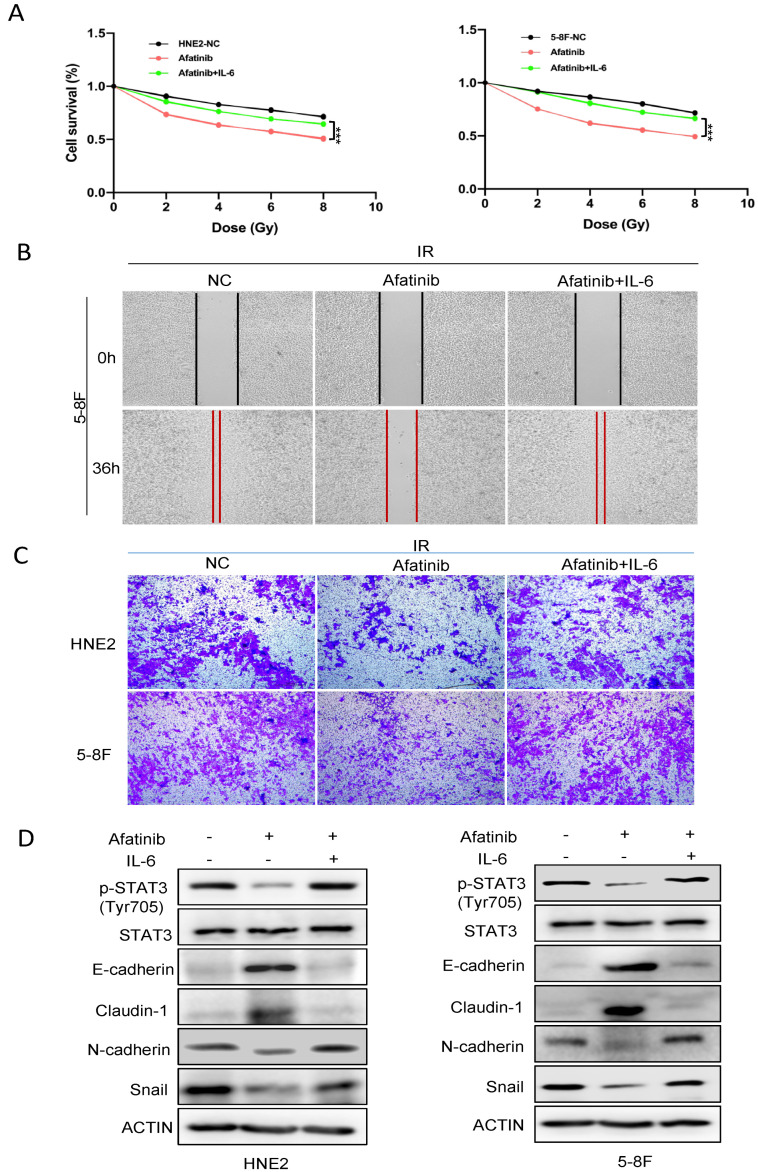
Methods: Quantitative phosphorylated proteomics and bioinformatics analysis were performed to illustrate the global phosphoprotein changes. The activity of the CD44-Stat3 axis and Epithelial-Mesenchymal Transition (EMT)-linked markers were evaluated by Western blotting. Wound healing and transwell assays were used to determine the levels of cell migration upon afatinib combined IR treatment. Cell proliferation was tested by CCK-8 assay. A pharmacological agonist by IL-6 was applied to activate Stat3. The xenograft mouse model was treated with afatinib, radiation or a combination of afatinib and radiation to detect the radiosensitivity of afatinib in vivo.

Results: In the present study, we discovered that afatinib triggered global protein phosphorylation alterations in NPC cells. Further, bioinformatics analysis indicated that afatinib inhibited the CD44-Stat3 signaling and subsequent EMT process. Moreover, functional assays demonstrated that afatinib combined radiation treatment remarkably impeded cell viability, migration, EMT process and CD44-Stat3 activity in vitro and in vivo. In addition, pharmacological stimulation of Stat3 rescued radiosensitivity and biological functions induced by afatinib in NPC cells. This suggested that afatinib reversed the EMT process by blocking the activity of the CD44-Stat3 axis.

Conclusion: Collectively, this work identifies the molecular mechanism of afatinib as a radiation sensitizer, thus providing a potentially useful combination treatment and drug target for NPC radiosensitization. Our findings describe a new function of afatinib in radiosensitivity and cancer treatment.

Keywords: CD44-Stat3 signaling pathway; afatinib; epithelial-to-mesenchymal transition (EMT); nasopharyngeal carcinoma (NPC); radiosensitivity.


Conflict of interest statement

The authors declare that they have no competing interests.

Figures

Figure 1
Figure 1
Schematic illustration of the TMT-based quantitative phosphoproteomic workflow. HNE2 cells pre-treated with IR or IR combined afatinib were conducted to TMT labeling, TiO2 enrichment and LC-MS/MS.
Figure 2
Figure 2
The distribution of phosphorylation sites. (A) Distribution of identified and quantified phosphorylated sites, phosphopeptides and phosphoproteins. (B) Distribution of phosphorylated sites on serine, threonine, and tyrosine. (C) Distribution of phosphorylated sites in a protein. (D) Volcano plots of group comparisons (Afatinib + IR vs. IR) showing the adjusted significance p-value (log2) versus fold change (log2). Red dots indicate significantly up-regulated phosphoproteins and blue dots indicate down-regulated.
Figure 3
Figure 3
Functional characteristics and KEGG analysis of DEPs. (A) The biological processes analysis of DEPs. (B) The cellular components analysis of DEPs. (C) The molecular functions analysis of DEPs. (D) KEGG pathway enrichment analysis of DEPs.
Figure 4
Figure 4
Phosphorylated proteomics analysis revealed influence on CD44-Stat3 axis. (A) Functions of the proteoglycans in cancer signaling pathway were presented. (B) DEPs interacted with CD44 and Stat3 were analyzed. (C,D) HNE2 cells were treated with afatinib or IR, WCLs were prepared at 24 h post-treatment and analyzed by immunoblotting with indicated antibodies (C). the statistical analysis is shown in (D). *** denotes p < 0.001. Error bars represent ±SD of triplicate experiments.
Figure 5
Figure 5
Afatinib combined IR treatment inhibited the EMT process. (A,B) 5-8F (A) and HNE2 cells (B) were treated as indicated and cell migration was measured by wound healing assay. (C) 5-8F and HNE2 cells were treated as indicated and cell migration was assessed by transwell assay without Matrigel. Cellular migration was plotted as the average number of cells in three different areas. (D) 5-8F and HNE2 cells were treated as indicated, WCLs were prepared at 24 h post-treatment and analyzed by immunoblotting with representative EMT marker antibodies. * denotes p < 0.05, ** denotes p < 0.01, *** denotes p < 0.001. Error bars represent ±SD of triplicate experiments.
Figure 6
Figure 6
Stat3 agonist reversed sensitivity to IR by regulating EMT. (A) HNE2 cells were treated with or without IL-6 after exposure to afatinib combined IR treatment and cell viability was evaluated by CCK-8 assay. (B,C) HNE2 and 5-8F cells were treated with or without IL-6, cell migration was measured by wound healing assay (B) and transwell assay (C). (D) HNE2 and 5-8F cells were treated with IL-6 after exposure to IR, WCLs were prepared and analyzed by immunoblotting with indicated antibodies. *** denotes p < 0.001.
Figure 7
Figure 7
Afatinib enhanced sensitivity to IR in vivo. (A) Schematic of the xenograft mouse experiment. 5-8F cells were injected subcutaneously into the right flank of male nude mice. When tumor volume reached about 200 mm3, mice were induced with indicated treatments. After two weeks, the mice were executed, HE and IHC experiments were performed. (B) Representative images of dissected xenografts. (C,D) Tumor volume (C) and tumor weight curve (D). (E) Representative images of HE and IHC staining from xenografts. * denotes p < 0.05.

References

    1. Sung H., Ferlay J., Siegel R.L., Laversanne M., Soerjomataram I., Jemal A., Bray F. Global Cancer Statistics 2020: GLOBOCAN Estimates of Incidence and Mortality Worldwide for 36 Cancers in 185 Countries. CA Cancer J. Clin. 2021;71:209–249. doi: 10.3322/caac.21660. - DOI - PubMed
    1. Tang L.-L., Chen W.-Q., Xue W.-Q., He Y.-Q., Zheng R.-S., Zeng Y.-X., Jia W.-H. Global trends in incidence and mortality of nasopharyngeal carcinoma. Cancer Lett. 2016;374:22–30. doi: 10.1016/j.canlet.2016.01.040. - DOI - PubMed
    1. Chen Y.P., Chan A.T.C., Le Q.T., Blanchard P., Sun Y., Ma J. Nasopharyngeal carcinoma. Lancet. 2019;394:64–80. doi: 10.1016/S0140-6736(19)30956-0. - DOI - PubMed
    1. Blanchard P., Lee A., Marguet S., Leclercq J., Ng W.T., Ma J., Chan A.T.C., Huang P.-Y., Benhamou E., Zhu G., et al. Chemotherapy and radiotherapy in nasopharyngeal carcinoma: An update of the MAC-NPC meta-analysis. Lancet Oncol. 2015;16:645–655. doi: 10.1016/S1470-2045(15)70126-9. - DOI - PubMed
    1. Luftig M. Heavy LIFting: Tumor promotion and radioresistance in NPC. J. Clin. Investig. 2013;123:4999–5001. doi: 10.1172/JCI73416. - DOI - PMC - PubMed

LinkOut - more resources