. 2023 Jan 9;16(1):100.
doi: 10.3390/ph16010100.

Momordica cochinchinensis (Gấc) Seed Extracts Induce Apoptosis and Necrosis in Melanoma Cells

Affiliations

Momordica cochinchinensis (Gấc) Seed Extracts Induce Apoptosis and Necrosis in Melanoma Cells

Dao Nguyen et al. Pharmaceuticals (Basel). .

Abstract

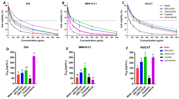
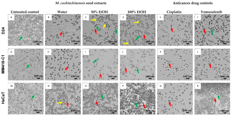
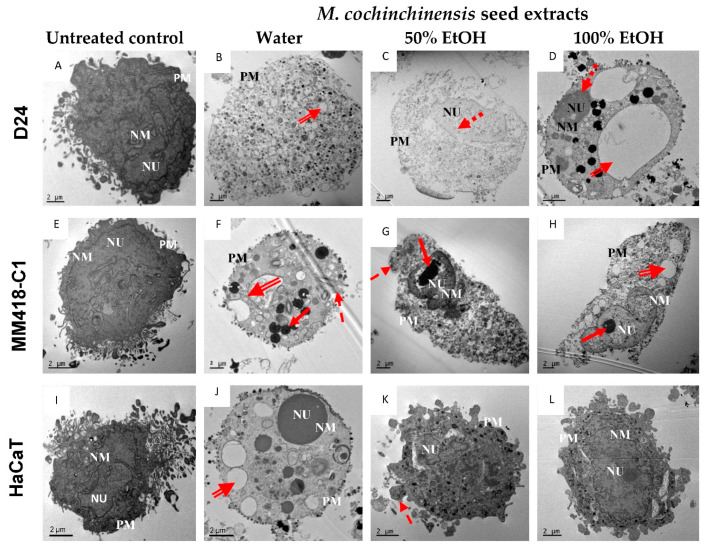
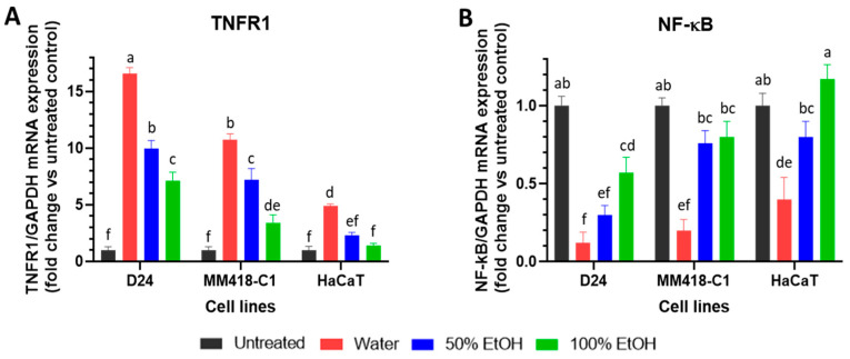
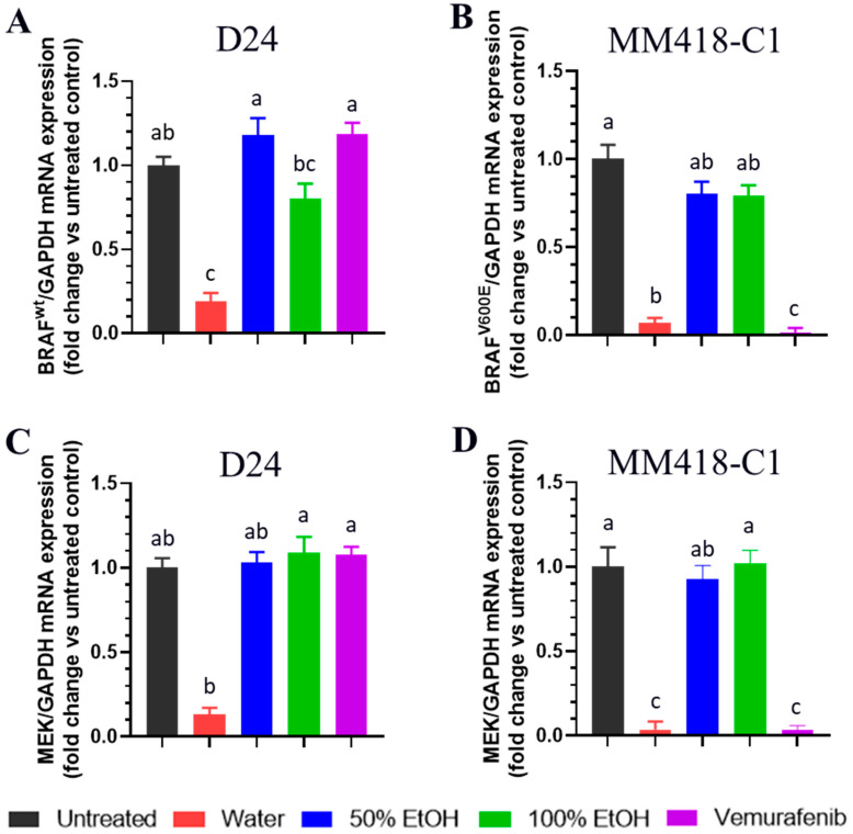
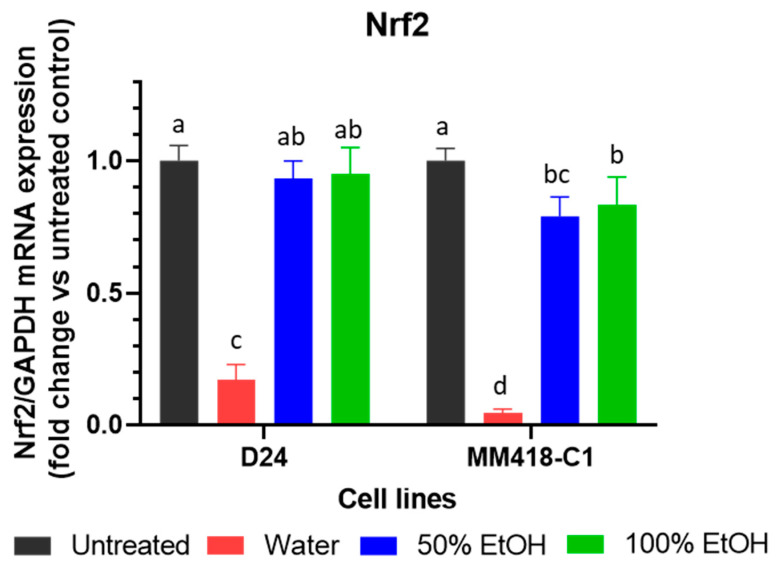
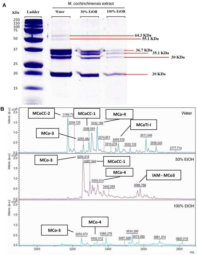
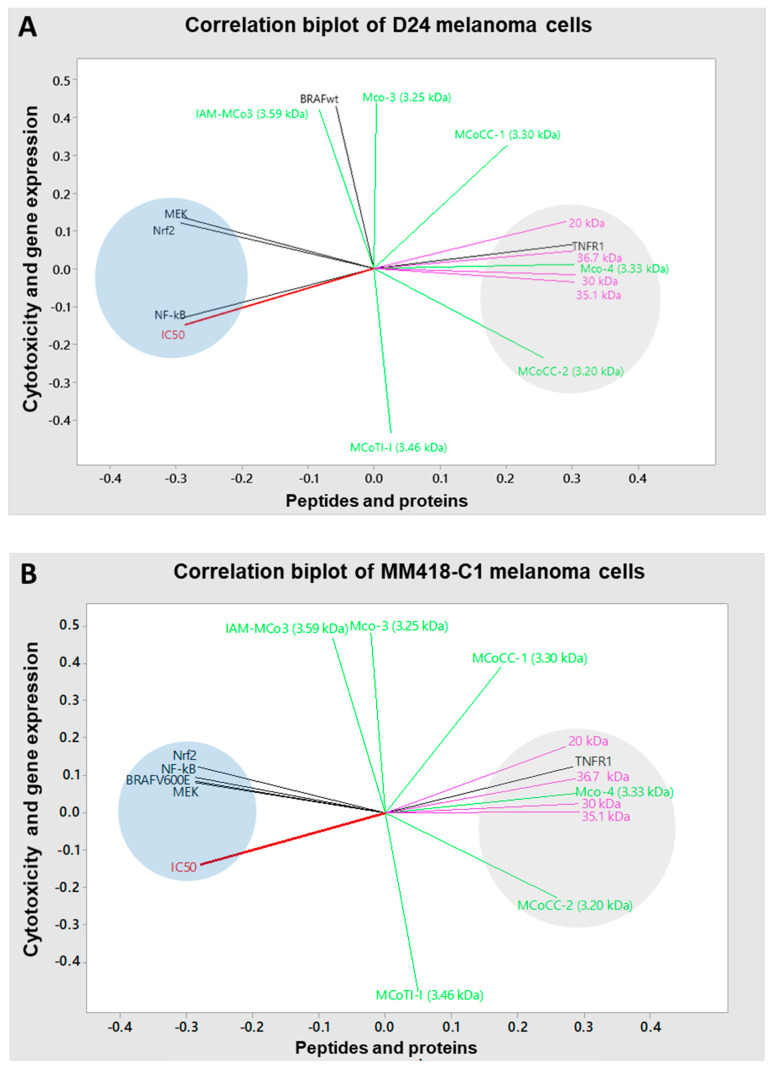
Momordica cochinchinensis is a herbal medicine used throughout Asia and this study investigated the antimelanoma potentials and molecular mechanisms of M. cochinchinensis seed with emphasis on extraction to optimise bioactivity. Overall, the aqueous extract was superior, with a wider diversity and higher concentration of proteins and peptides that was more cytotoxic to the melanoma cells than other extraction solvents. The IC50 of the aqueous extract on melanoma cells were similar to treatment with current anticancer drugs, vemurafenib and cisplatin. This cytotoxicity was cancer-specific with lower cytotoxic effects on HaCaT epidermal keratinocytes. Cytotoxicity correlated with MAPK signalling pathways leading to apoptosis and necrosis induced by triggering tumour necrosis factor receptor-1 (TNFR1), reducing the expression of nuclear factor kappa B (NF-kB), and suppression of BRAF/MEK. This efficacy of M. cochinchinensis seed extracts on melanoma cells provides a platform for future clinical trials as potent adjunctive therapy for metastatic melanoma.

Keywords: BRAF oncogene; M. cochinchinensis; MAPK signalling pathway; NF-kB; Nrf2; TNFR1; cancer; gac fruit; melanoma.


Conflict of interest statement

The authors declare no competing interest.

Figures

Figure 1
Figure 1
Effect of M. cochinchinensis seed extracts on the cell viability and half inhibition concentration (IC50) of D24 and MM418-C1 melanoma and HaCaT cells (n = 3). Dose response of cells (% viability) after 48 h treatment with different concentrations of water, 50% EtOH and 100% EtOH extracts, cisplatin and vemurafenib on D24 melanoma cells (A), MM418-C1 melanoma cells (B), and HaCaT cells (C). Cell viability (%) was expressed as the average ratio of the treated divided by the untreated control (the dotted lines represented 50% cell viability); half inhibition concentration (IC50) of the seed extracts for D24 melanoma cells (D), MM418-C1 melanoma cells (E), and HaCaT cells (F). The results were presented as the mean ± SEM, different letters indicated statistical significance at p ≤ 0.05 between different treatments (Analysis by One-way ANOVA with Turkey’s LSD test).
Figure 2
Figure 2
Effect of 48 h exposure to 100 µg/mL M. cochinchinensis seed extracts on melanoma (D24 and MM418-C1) and HaCaT cells compared to cisplatin and vemurafenib treatment. Green arrows indicate normal cells, red arrows indicate cell shrinkage, yellow arrows indicate cell swelling (Scale bar = 100 µm). (A): Untreated D24 cells (control), (B): D24 cells treated with water extract, (C): D24 cells treated with 50% EtOH extract, (D): D24 cells treated with 100% EtOH extract, (E): D24 cells treated with cisplatin, (F): D24 cells treated with vemurafenib, (G): Untreated MM418-C1 cells (control), (H): MM418-C1 cells treated with water extract, (I): MM418-C1 cells treated with 50% EtOH extract, (J): MM418-C1 cells treated with 100% EtOH extract, (K): MM418-C1 cells treated with cisplatin, (L): MM418-C1 cells treated with vemurafenib, (M): Untreated HaCaT cells (control), (N): HaCaT cells treated with water extract, (O): HaCaT cells treated with 50% EtOH extract, (P): HaCaT cells treated with 100% EtOH extract, (Q): HaCaT cells treated with cisplatin, (R): HaCaT cells treated with vemurafenib.
Figure 3
Figure 3
Effect of M. cochinchinensis seed extracts on the morphology of melanoma (D24 and MM418-C1) and HaCaT cells. The cells were treated with 100 µg/mL of the seed extracts for 48 h and examined under electron microscopy. Solid-line arrow: chromatin condensation, dotted-line arrow: margination of chromatin, hatched-line arrow: cell membrane blebbing, double-line arrow: vacuolisation, NU: nucleus, NM: nuclear membrane, PM: plasma membrane. (A): Untreated D24 cells (control), (B): D24 cells treated with water extract, (C): D24 cells treated with 50% EtOH extract, (D): D24 cells treated with 100% EtOH extract, (E): Untreated MM418-C1 cells (control), (F): MM418-C1 cells treated with water extract, (G): MM418-C1 cells treated with 50% EtOH extract, (H): MM418-C1 cells treated with 100% EtOH extract, (I): Untreated HaCaT cells (control), (J): HaCaT cells treated with water extract, (K): HaCaT cells treated with 50% EtOH extract, (L): HaCaT cells treated with 100% EtOH extract.
Figure 4
Figure 4
Effect of M. cochinchinensis seed extracts on the expression of. TNFR1 (A) and NF-kB (B) mRNA in D24 and MM418-C1 melanoma and HaCaT cells. The cells were treated with 100 µg/mL of the different seed extracts for 48 h prior to RNA collection. Results were presented as the fold change compared to untreated controls ± SEM (n = 3), different letters indicated statistical significance at p ≤ 0.05 between different treatments (Analysis by One-way ANOVA with Turkey’s LSD test).
Figure 5
Figure 5
Effect of M. cochinchinensis seed extracts on the expression of BRAFWT gene in D24 (A), BRAFV600E gene in MM418-C1 (B), and MEK1 genes in D24 (C) and MM418-C1 (D) melanoma cells. The cells were treated with 100 µg/mL of the different seed extracts or vemurafenib for 48 h prior to RNA collection. Results were presented as the fold change compared to untreated controls ± SE, different letters indicated statistical significance at p ≤ 0.05 between different treatments (Analysis by One-way ANOVA with Turkey’s LSD test).
Figure 6
Figure 6
Effect of M. cochinchinensis seed extracts on the expression of Nrf2 mRNA in D24 and MM418-C1 melanoma cells. The cells were treated with 100 µg/mL of the different seed extracts for 48 h prior to RNA collection. Results were presented as the fold change compared to untreated controls ± SE, different letters indicated statistical significance at p ≤ 0.05 between different treatments (Analysis by One-way ANOVA with Turkey’s LSD test).
Figure 7
Figure 7
Protein (A) and peptide (B) profiles of M. cochinchinensis seed extracts.
Figure 8
Figure 8
Correlation biplot of cytotoxicity (IC50) and gene expression with M. cochinchinensis seed proteins and peptides in D24 (A) and MM418-C1 melanoma cells (B) constructed using the principal component analysis method (PCA) with Minitab statistical software (Version 18). Red = IC50, black = gene expressions, purple = proteins and green = peptides.

Similar articles

Cited by

References

    1. Sung H., Ferlay J., Siegel R.L., Laversanne M., Soerjomataram I., Jemal A., Bray F. Global Cancer Statistics 2020: GLOBOCAN Estimates of Incidence and Mortality Worldwide for 36 Cancers in 185 Countries. CA Cancer J. Clin. 2021;71:209–249. doi: 10.3322/caac.21660. - DOI - PubMed
    1. Ottaviano M., Giunta E., Tortora M., Curvietto M., Attademo L., Bosso D., Cardalesi C., Rosanova M., de Placido P., Pietroluongo E., et al. BRAF Gene and Melanoma: Back to the Future. Int. J. Mol. Sci. 2021;22:3474. doi: 10.3390/ijms22073474. - DOI - PMC - PubMed
    1. Malissen N., Grob J.-J. Metastatic Melanoma: Recent Therapeutic Progress and Future Perspectives. Drugs. 2018;78:1197–1209. doi: 10.1007/s40265-018-0945-z. - DOI - PubMed
    1. Berthenet K., Ferrer C.C., Fanfone D., Popgeorgiev N., Neves D., Bertolino P., Gibert B., Hernandez-Vargas H., Ichim G. Failed Apoptosis Enhances Melanoma Cancer Cell Aggressiveness. Cell Rep. 2020;31:107731. doi: 10.1016/j.celrep.2020.107731. - DOI - PubMed
    1. Chan X.Y., Singh A., Osman N., Piva T.J. Role Played by Signalling Pathways in Overcoming BRAF Inhibitor Resistance in Melanoma. Int. J. Mol. Sci. 2017;18:1527. doi: 10.3390/ijms18071527. - DOI - PMC - PubMed