. 2022 Dec 22;15(1):35.
doi: 10.3390/pharmaceutics15010035.

Antifungal Combination Eye Drops for Fungal Keratitis Treatment

Affiliations

Antifungal Combination Eye Drops for Fungal Keratitis Treatment

Victoria Díaz-Tomé et al. Pharmaceutics. .

Abstract

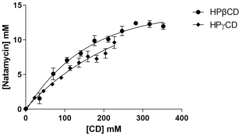
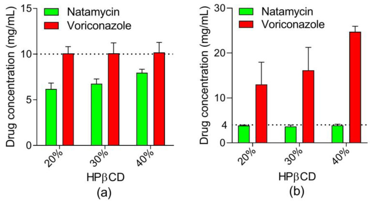
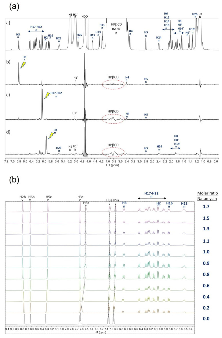
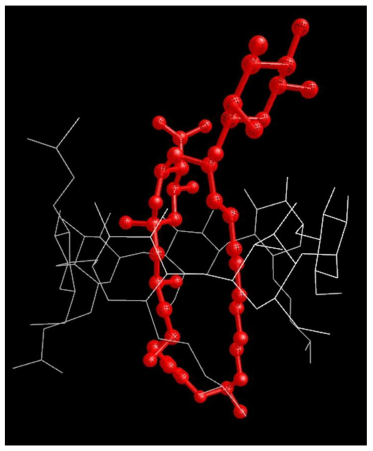
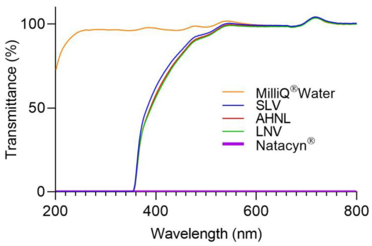
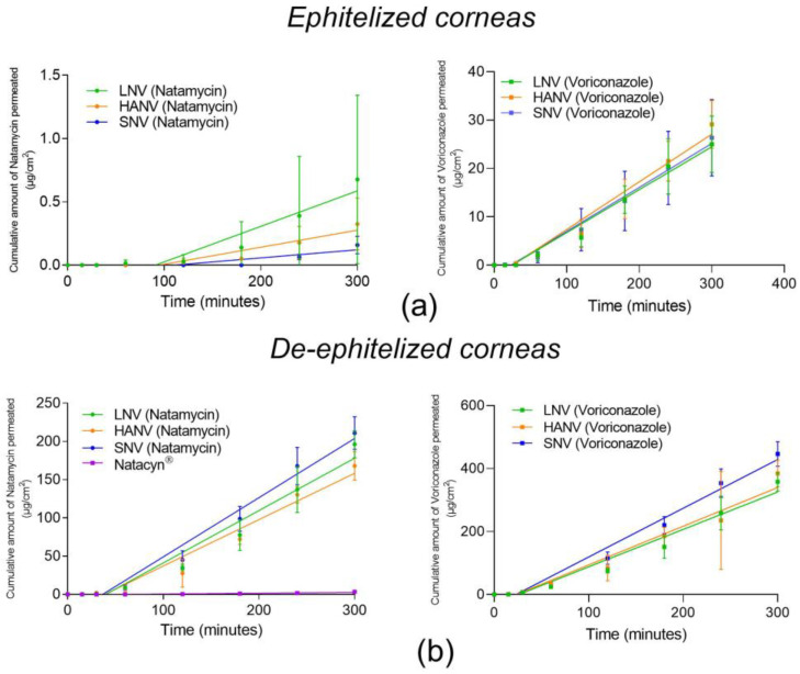
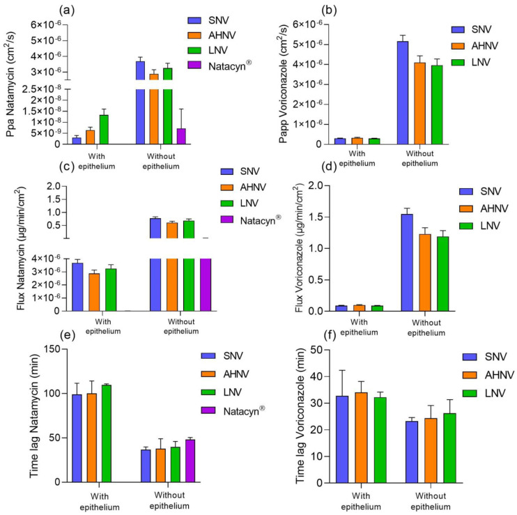
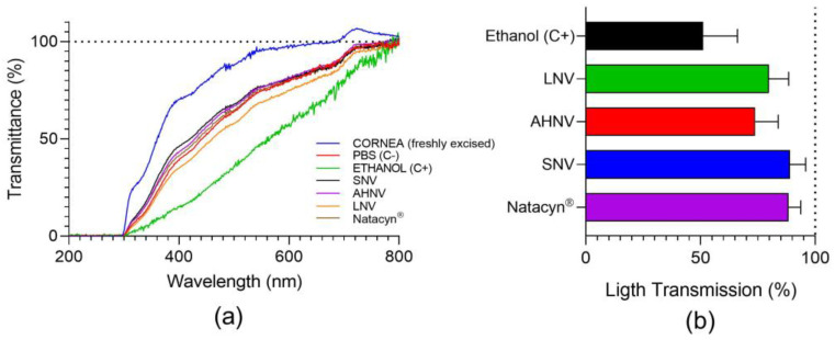
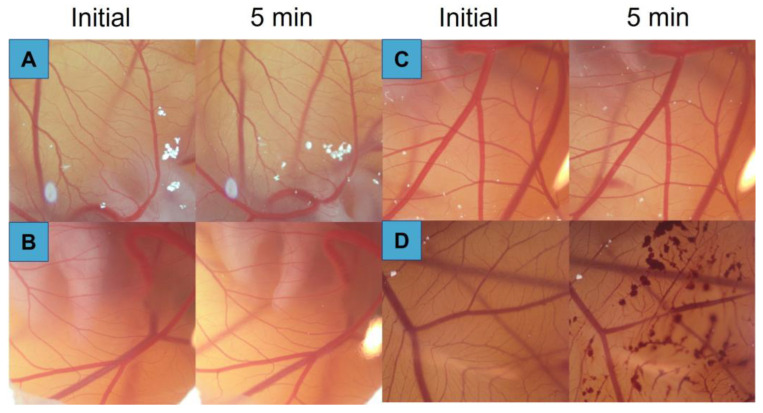
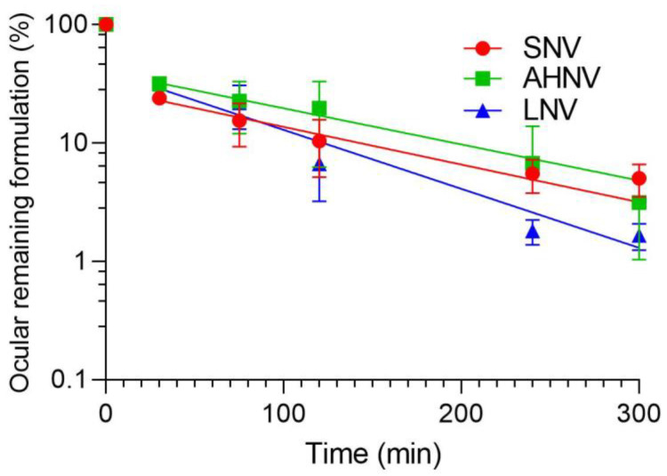
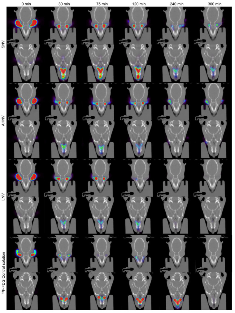
Fungal keratitis (FK) is a corneal mycotic infection that can lead to vision loss. Furthermore, the severity of FK is aggravated by the emergence of resistant fungal species. There is currently only one FDA-approved formulation for FK treatment forcing hospital pharmacy departments to reformulate intravenous drug preparations with unknown ocular bioavailability and toxicity. In the present study, natamycin/voriconazole formulations were developed and characterized to improve natamycin solubility, permanence, and safety. The solubility of natamycin was studied in the presence of two cyclodextrins: HPβCD and HPγCD. The HPβCD was chosen based on the solubility results. Natamycin/cyclodextrin (HPβCD) inclusion complexes characterization and a competition study between natamycin and voriconazole were conducted by NMR (Nuclear Magnetic Resonance). Based on these results, several eye drops with different polymer compositions were developed and subsequently characterized. Permeability studies suggested that the formulations improved the passage of natamycin through the cornea compared to the commercial formulation Natacyn®. The ocular safety of the formulations was determined by BCOP and HET-CAM. The antifungal activity assay demonstrated the ability of our formulations to inhibit the in vitro growth of different fungal species. All these results concluded that the formulations developed in the present study could significantly improve the treatment of FK.

Keywords: PET/CT imaging; cyclodextrin; cyclodextrin aggregates; fungal keratitis; natamycin; nuclear magnetic resonance; voriconazole.


Conflict of interest statement

The authors declare no conflict of interest. The funders had no role in the design of the study; in the collection, analyses, or interpretation of data; in the writing of the manuscript; or in the decision to publish the results.

Figures

Figure 1
Figure 1
Phase solubility diagrams for natamycin, obtained with 2 different types of cyclodextrin (HPβCD and HPγCD) at 25 °C in water (mean ± SD, n = 6).
Figure 2
Figure 2
Transmission Electron Microscopy (TEM) images of saturated solution of (a) natamycin in 40% (w/v) HPβCD solution and 1% (w/v) voriconazole solution and (b) saturated solution of natamycin in 40% (w/v) HPβCD solution.
Figure 3
Figure 3
(a) Voriconazole concentration obtained in presence of 4 mg/mL of natamycin and different concentrations of HPβCD. (b) Natamycin concentration obtained in presence of 10 mg/mL of voriconazole and different concentrations of HPβCD.
Figure 4
Figure 4
(a) NMR spectra of natamycin:HPβCD 1:1 in D2O showing the assignment of signals of natamycin (n) and HPβCD (h). (a) 1H reference spectrum. (b) STDoff-on spectrum with on-saturation at 6.79 ppm (H-3 signal of n). (c) STDoff-on with on-saturation at 6.05 ppm (H-17 to H-22 signal of n). (d) STDoff-on with on-saturation at 5.91 ppm (H-2 signal of n). The atom numbering used to identify the signals of voriconazole follows S1. (b) NMR titration competition assay with natamycin at a constant molar ratio voriconazole:HPβCD 1:1. Stack of spectra showing the aromatic region of the 1H-NMR spectrum during the titration. The atom numbering used to identify voriconazole (v) and natamycin (n) follows S1. Stripped lines were drawn to guide the eye for the changes in chemical shift (i.e., CSPs) of certain signals of voriconazole.
Figure 5
Figure 5
Optimized molecular mechanics model of the natamycin complex with HPβCD. Red lines represent natamycin and white lines represent HPβCD.
Figure 6
Figure 6
Scan light transmittance from 200 to 800 nm for the formulations including natamycin. MilliQ® water was used as a transparent formulation in all light ranges.
Figure 7
Figure 7
In vitro release profiles of natamycin/voriconazole formulations. All data were fitted to zero-order kinetics.
Figure 8
Figure 8
Cumulative amount of drug permeated (µg/cm2) through epithelized (a) bovine and de-epithelized (b) corneas for natamycin (left) and voriconazole (right).
Figure 9
Figure 9
(a) Apparent permeability (Papp) of natamycin, (b) apparent permeability (Papp) of voriconazole, (c) flux of natamycin, (d) flux of voriconazole, (e) lag time of natamycin, and (f) lag time of voriconazole of natamycin and voriconazole formulations across epithelized and deepithelized bovine corneas. * Natacyn® permeability values are not shown because there was no permeability of natamycin from this formulation.
Figure 10
Figure 10
(a) Transmittance values in the ultra-visible light spectrum (200–800 nm) of bovine corneas treated 10 min with SNV, AHNV, and LNV. Values are compared with ethanol (C+: positive control), PBS (C−: negative control), and untreated corneas. (b) Transmitted light (%) (opacity) values of bovine corneas treated with SNV, AHNV, and LNV. Data were compared with ethanol (C+: positive control).
Figure 11
Figure 11
5 min post-instillation images of Hen´s egg test on the chorioallantoic membrane (HET-CAM) for different natamycin/voriconazole formulations. (A) SNV; (B) AHNV; (C) LNV; (D) NaOH 0.1 M.
Figure 12
Figure 12
SNV, AHNV, and LNV clearance ratio from the ocular surface determination by PET. Ratio CT/Cinitial was calculated assuming Cinitial value obtained in the Regions of Interest (ROI).
Figure 13
Figure 13
Coronal PET/CT images of rat eyes treated with SNV, AHNV, and LNV over time. Data were compared with a 18F-FDG control solution. The amount of formulation on the ocular surface is coded on a color scale: blue areas show a low radioactive activity; red areas show high radioactive activity.

References

    1. Rai M., Occhiutto M.L. Mycotic Keratitis. CRC Press; Boca Raton, FL, USA: 2019.
    1. Taechajongjintana M., Kasetsuwan N., Reinprayoon U., Sawanwattanakul S., Pisuchpen P. Effectiveness of voriconazole and corneal cross-linking on Phialophora verrucosa keratitis: A case report. J. Med. Case Rep. 2018;12:225. doi: 10.1186/s13256-018-1765-1. - DOI - PMC - PubMed
    1. Bourcier T., Sauer A., Dory A., Denis J., Sabou M. Fungal keratitis. J. Fr. Ophtalmol. 2017;40:e307–e313. doi: 10.1016/j.jfo.2017.08.001. - DOI - PubMed
    1. Lund O.E., Miño de Kaspar H., Klauss V. Strategy for examination and therapy of mycotic keratitis. Klin. Monbl. Augenheilkd. 1993;202:188–194. doi: 10.1055/s-2008-1045581. - DOI - PubMed
    1. Saha S., Banerjee D., Khetan A., Sengupta J. Epidemiological profile of fungal keratitis in urban population of west Bengal, India. Oman. J. Ophthalmol. 2009;2:114–118. doi: 10.4103/0974-620X.57310. - DOI - PMC - PubMed

LinkOut - more resources