. 2022 Dec 28;15(1):96.
doi: 10.3390/pharmaceutics15010096.

A Supramolecular Nanoassembly of Lenvatinib and a Green Light-Activatable NO Releaser for Combined Chemo-Phototherapy

Affiliations

A Supramolecular Nanoassembly of Lenvatinib and a Green Light-Activatable NO Releaser for Combined Chemo-Phototherapy

Francesca Laneri et al. Pharmaceutics. .

Abstract

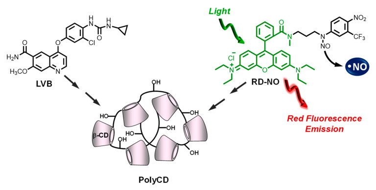
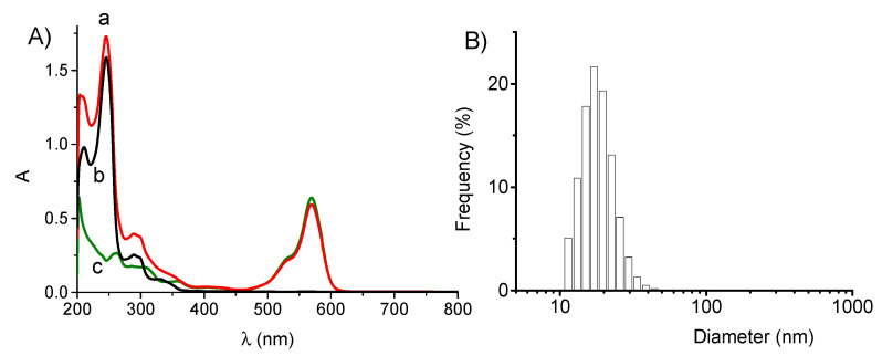
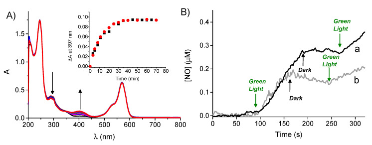
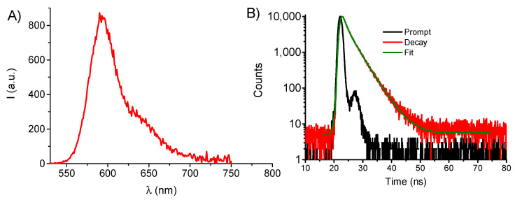
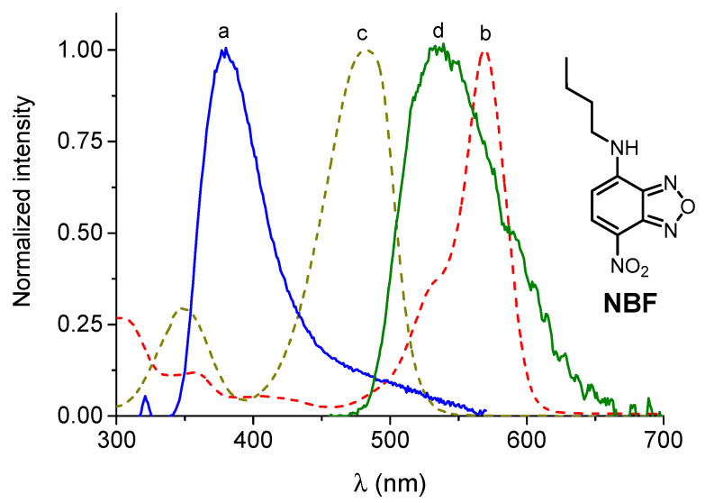
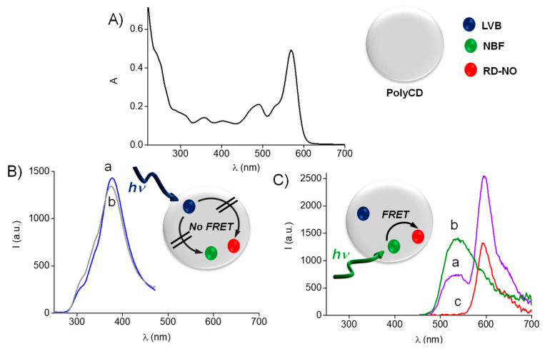
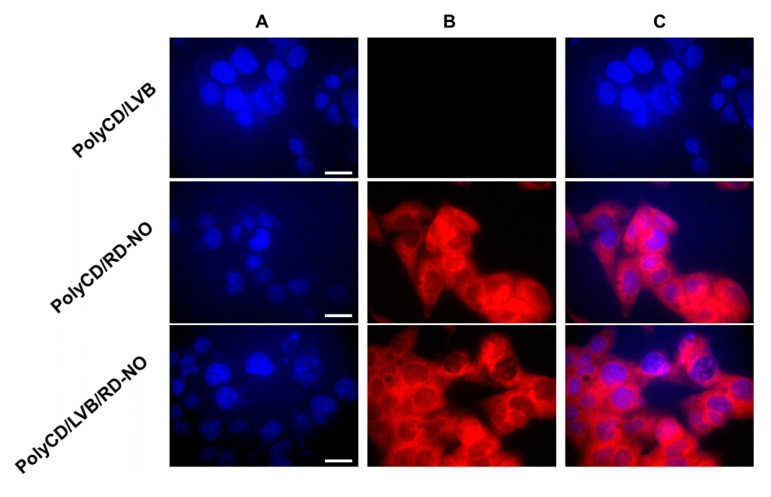
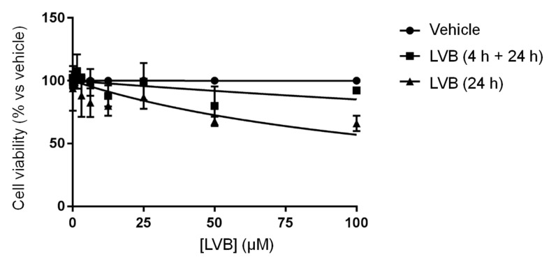
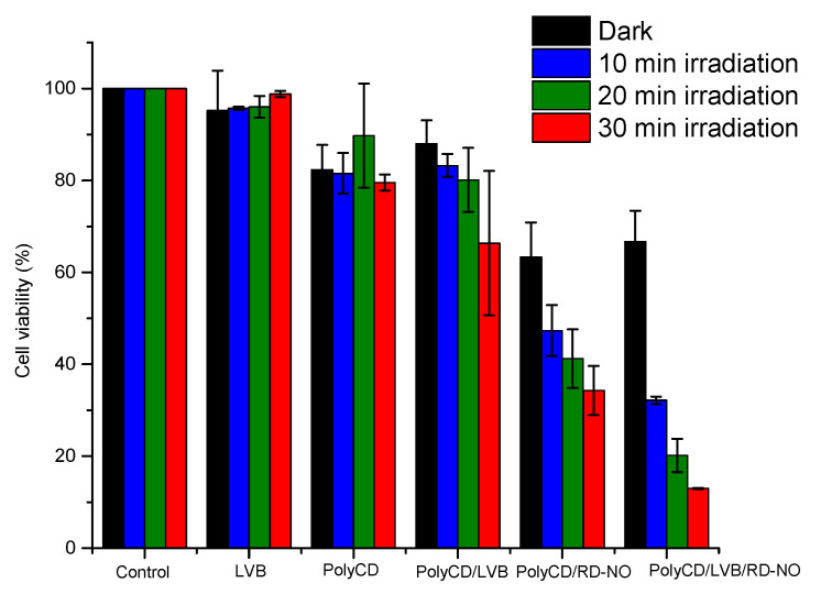
The chemotherapeutic Lenvatinib (LVB) and a nitric oxide (NO) photodonor based on a rhodamine antenna (RD-NO) activatable by the highly compatible green light are supramolecularly assembled by a β-cyclodextrin branched polymer (PolyCD). The poorly water-soluble LVB and RD-NO solubilize very well within the polymeric host leading to a ternary supramolecular nanoassembly with a diameter of ~55 nm. The efficiency of the NO photorelease and the typical red fluorescence of RD-NO significantly enhance within the polymer due to its active role in the photochemical and photophysical deactivation pathways. The co-presence of LVB within the same host does not affect either the nature or the efficiency of the photoinduced processes of RD-NO. Besides, irradiation of RD-NO does not lead to the decomposition of LVB, ruling out any intermolecular photoinduced process between the two guests despite sharing the same host. Ad-hoc devised Förster Resonance Energy Transfer experiments demonstrate this to be the result of the not close proximity of the two guests, which are confined in different compartments of the same polymeric host. The supramolecular complex is stable in a culture medium, and its biological activity has been evaluated against HEP-G2 hepatocarcinoma cell lines in the dark and under irradiation with visible green light, using LVB at a concentration well below the IC50. Comparative experiments performed using the polymeric host encapsulating the individual LVB and RD-NO components under the same experimental conditions show that the moderate cell mortality induced by the ternary complex in the dark increases significantly upon irradiation with visible green light, more likely as the result of synergism between the NO photogenerated and the chemotherapeutic.

Keywords: chemotherapeutic; combination therapy; cyclodextrin polymers; light; nitric oxide.


Conflict of interest statement

The authors declare no conflict of interest.

Figures

Scheme 1
Scheme 1
Molecular structures of the guests LVB and RD-NO, which are supramolecularly assembled in the branched polymeric host PolyCD.
Figure 1
Figure 1
(A) Absorption spectrum of LVB in PBS (pH 7.4) at 4.1 µM (a) and in the presence of PolyCD (2 mg mL−1) at 25 µM (b), 40 µM (c), 50 µM (d) and 60 µM I (e). The spectrum of a 60 µM LVB solution in methanol is shown for the sake of comparison (f). (B) Absorbance value at 245 nm of LVB at different concentrations in the presence of PolyCD. T = 25 °C; cell path = 0.1 cm.
Figure 2
Figure 2
(A) Absorption spectra of PolyCD (2 mg mL−1) in PBS (pH 7.4) co-loaded with LVB (25 µM) and RD-NO (6 µM) (a) and, for the sake of comparison, loaded only with LVB (b) and RD-NO (c). T = 25 °C; cell path = 1 cm. (B) Hydrodynamic diameter of sample a in Figure 2A.
Figure 3
Figure 3
(A) Absorption spectral changes observed upon 532 nm light irradiation of a PBS (pH 7.4) solution of PolyCD (2 mg mL−1) co-loaded with LVB (25 µM) and RD-NO (6 µM). The inset shows the absorbance changes at 397 nm of PolyCD (2 mg mL−1) co-loaded with LVB (25 µM) and RD-NO (6 µM) (λ) and, for comparison, the same host in the absence of LVB (ν). (B) NO release profile observed for PolyCD (2 mg mL−1) co-loaded with LVB (25 µM) and RD-NO (6 µM) (a) and, for comparison, the same host in the absence of LVB (b). T = 25 °C.
Figure 4
Figure 4
(A) Fluorescence emission spectrum observed at λexc = 520 nm light excitation for a PBS (pH 7.4) solution of PolyCD (2 mg mL−1) co-loaded with LVB (25 µM) and RD-NO (6 µM). (B) Fluorescence decay and the related fitting of the same solution recorded at λexc = 455 nm and λem = 590 nm. T = 25 °C.
Figure 5
Figure 5
Normalized fluorescence spectra of LVB (a) (λexc = 240 nm), NBF (d) (λexc = 440 nm), and absorption spectra of NBF (c) and RD-NO (b). All guests are loaded in PolyCD (2 mg mL−1) dissolved in PBS (pH 7.4). T = 25 °C.
Figure 6
Figure 6
(A) Absorption spectrum of PolyCD (2 mg mL−1) in PBS (pH 7.4) co-loaded with LVB, RD-NO, and NBF (5 µM). (B) Fluorescence emission spectra of the sample as in (A) (a) and PolyCD (2 mg mL−1) in PBS (pH 7.4) loaded only with LVB at the same concentration (b) recorded at λexc = 240 nm. (C) Fluorescence emission spectra of the sample as in (A) (a) and PolyCD (2 mg mL−1) in PBS (pH 7.4) loaded only with NBF (b) and RD-NO (c) recorded at λexc = 445 nm.
Figure 7
Figure 7
Fluorescence microscopy analysis of HEP-G2 hepatocarcinoma cell lines treated with PolyCD (2 mg mL−1) in PBS (pH 7.4) loaded with LVB (25 µM), RD-NO (6 µM) and both components and stained with DAPI. The cells were analyzed with a DAPI emission filter (A), a rhodamine emission filter (B), or by merging images (A,B) (C). Scale bar = 50 µM.
Figure 8
Figure 8
Viability of HEP-G2 hepatocarcinoma cells as a function of the concentration of free LVB.
Figure 9
Figure 9
Cell viability was observed 24 h after incubating HEP-G2 hepatocarcinoma cells with free LVB, PolyCD, and the supramolecular complexes PolyCD/LVB, PolyCD/RD-NO, and PolyCD/LVB/RD-NO in the dark and upon different irradiation times (occurred after the first 4 h of incubation) at λexc > 500 nm. [LVB] = 50 µM; [RD-NO] = 6 µM; [PolyCD] = 2 mg mL−1.

References

    1. Schlumberger M., Tahara M., Wirth L.J., Robinson B., Brose M.S., Elisei R., Habra M.A., Newbold K., Shah M.H., Hoff A.O., et al. Lenvatinib versus placebo in radioiodine-refractory thyroid cancer. N. Engl. J. Med. 2015;372:621–630. doi: 10.1056/NEJMoa1406470. - DOI - PubMed
    1. Kiyota N., Schlumberger M., Muro K., Ando Y., Takahashi S., Kawai Y., Wirth L., Robinson B., Sherman S., Suzuki T., et al. Subgroup analysis of Japanese patients in a phase 3 study of lenvatinib in radioiodine-refractory differentiated thyroid cancer. Cancer Sci. 2015;106:1714–1721. doi: 10.1111/cas.12826. - DOI - PMC - PubMed
    1. Al-Salama Z.T., Syed Y.Y., Scott L.J. Lenvatinib: A Review in Hepatocellular Carcinoma. Drugs. 2019;79:665–674. doi: 10.1007/s40265-019-01116-x. - DOI - PubMed
    1. Cabanillas M.E., Habra M.A. Lenvatinib: Role in thyroid cancer and other solid tumors. Cancer Treat. Rev. 2016;42:47–55. doi: 10.1016/j.ctrv.2015.11.003. - DOI - PubMed
    1. Kudo M., Finn R.S., Qin S., Han K.H., Ikeda K., Piscaglia F., Baron A., Park J.W., Han G., Jassem J., et al. Levantinib versus sorafenib in first-line treatment of patients with unresectable hepatocellular carcinoma: A randomised phase 3 non-inferiority trial. Lancet. 2018;391:1163–1173. doi: 10.1016/S0140-6736(18)30207-1. - DOI - PubMed

LinkOut - more resources