. 2023 Jan 10;12(2):330.
doi: 10.3390/plants12020330.

ATANN3 Is Involved in Extracellular ATP-Regulated Auxin Distribution in Arabidopsis thaliana Seedlings

Affiliations

ATANN3 Is Involved in Extracellular ATP-Regulated Auxin Distribution in Arabidopsis thaliana Seedlings

Jiawei Xu et al. Plants (Basel). .

Abstract

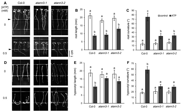
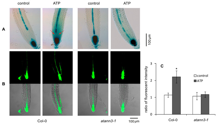
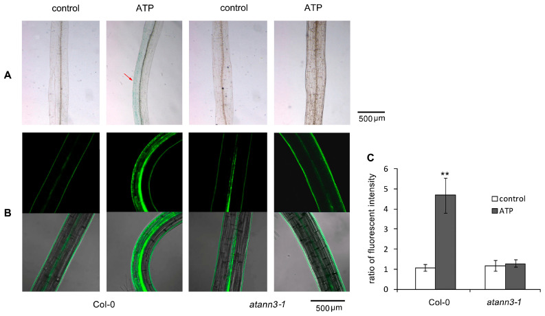
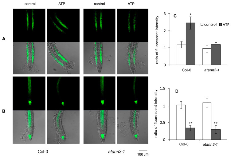
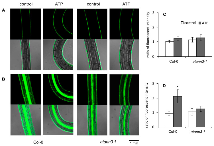
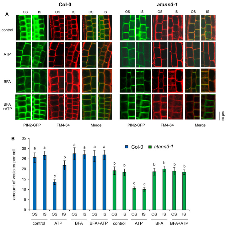
Extracellular ATP (eATP) plays multiple roles in plant growth and development, and stress responses. It has been revealed that eATP suppresses growth and alters the growth orientation of the root and hypocotyl of Arabidopsis thaliana by affecting auxin transport and localization in these organs. However, the mechanism of the eATP-stimulated auxin distribution remains elusive. Annexins are involved in multiple aspects of plant cellular metabolism, while their role in response to apoplastic signals remains unclear. Here, by using the loss-of-function mutations, we investigated the role of AtANN3 in the eATP-regulated root and hypocotyl growth. Firstly, the inhibitory effects of eATP on root and hypocotyl elongation were weakened or impaired in the AtANN3 null mutants (atann3-1 and atann3-2). Meanwhile, the distribution of DR5-GUS and DR5-GFP indicated that the eATP-induced asymmetric distribution of auxin in the root tips or hypocotyl cells occurred in wild-type control plants, while in atann3-1 mutant seedlings, it was not observed. Further, the eATP-induced asymmetric distribution of PIN2-GFP in root-tip cells or that of PIN3-GFP in hypocotyl cells was reduced in atann3-1 seedlings. Finally, the eATP-induced asymmetric distribution of cytoplasmic vesicles in root-tip cells was impaired in atann3-1 seedlings. Based on these results, we suggest that AtANN3 may be involved in eATP-regulated seedling growth by regulating the distribution of auxin and auxin transporters in vegetative organs.

Keywords: Arabidopsis thaliana; AtANN3; auxin; extracellular ATP (eATP); seedling growth.


Conflict of interest statement

The authors declare no conflict of interest.

Figures

Figure 1
Figure 1
AtANN3 is involved in the eATP-regulated seedling growth of Arabidopsis thaliana. (AC) The 4-day-old seedlings were transplanted onto the combined medium and cultured under light conditions for 5 more days. The concentration of ATP in the lower compartment was 0.5 mM. The triangles mark the border between the two growth media. (A) The growing seedlings and (B,C) the root length and root curvature of seedlings, respectively. (DF) Seeds were sown and cultured on the medium containing 0.5 mM ATP in the dark for 5 days. (D) The growing etiolated seedlings and (E,F) the hypocotyl length and curvature of seedlings, respectively. The scale bar is shown in the lower-right corner of (A). In each experiment, at least 30 seedlings were measured, and data obtained from at least three replicates were combined to obtain mean ± SD and then analyzed statistically. The same letter on the top of the columns indicates that means of different treatment groups were not significantly different based on Duncan’s multiple range test (p < 0.05).
Figure 2
Figure 2
AtANN3 is involved in the eATP-induced distribution of DR5-GUS or DR5-GFP in root-tip cells. The 4-day-old seedlings of DR5-GUS or DR5-GFP transgenic lines were transplanted onto the medium containing 0.5 mM ATP and cultured for 2 more days. (A) The DR5-GUS expression in representative root-tip cells; red arrows indicate the area with the accumulation of DR5-GUS. (B) The DR5-GFP fluorescence in representative root-tip cells. The scale bar is shown in the lower-right corner of the figure. (C) The fluorescence intensity ratio (FIR) in the cells at the inner side/outer side of the root curve. In this experiment, at least 15 samples were measured, and data obtained from at least three replicates were combined to obtain mean ± SD based on Student’s t-test (* p < 0.05).
Figure 3
Figure 3
AtANN3 is involved in the eATP-induced DR5-GUS or DR5-GFP distribution in the etiolated hypocotyl cells. The 4-day-old etiolated seedlings of DR5-GUS or DR5-GFP transgenic lines were transplanted onto the medium containing 0.5 mM ATP and cultured for 2 more days. (A) The DR5-GUS expression in representative hypocotyls. The red arrow indicates the accumulation area of DR5-GUS. (B) The DR5-GFP fluorescence in representative hypocotyls. The scale bar is shown in the lower-right corner of each figure. (C) The fluorescence intensity ratio (FIR) in cells at the inner side/outer side at the hypocotyl curve. In this experiment, at least 15 samples were measured, and data from at least three replicates were combined to obtain mean ±SD based on Student’s t-test p-values (** p < 0.01).
Figure 4
Figure 4
AtANN3 is involved in the eATP-induced PIN2-GFP distribution in root cells. The 4-day-old seedlings of PIN2-GFP or PIN3-GFP transgenic lines were transplanted onto the medium containing 0.5 mM ATP and cultured for 2 more days. (A,B) Fluorescent images of PIN2-GFP and PIN3-GFP in root tips, respectively. The scale bar is shown in the lower-right corner of the image. (C) The fluorescence intensity ratio of PIN2-GFP in cells at the inner side/outer side at the root curve. (D) The fluorescence intensity ratio of PIN3-GFP in QC and stele cells before and after the ATP treatment. The Fluorescence intensity in root tips of untreated Col-0 seedlings was set at 1. In each experiment, at least 15 samples were measured and data from at least three replicates were combined to obtain mean ± SD based on Student’s t-test p-values (* p < 0.05, ** p < 0.01).
Figure 5
Figure 5
AtANN3 is involved in the eATP-induced PIN3-GFP distribution in hypocotyl cells. The 4-day-old seedlings of PIN2-GFP or PIN3-GFP transgenic lines were transplanted onto the medium containing 0.5 mM ATP and cultured for 2 more days. (A,B) Fluorescence in the hypocotyl of PIN2-GFP (A) and PIN3-GFP (B) transformant seedlings. The scale bar is shown in the lower-right corner of the image. (C,D) The fluorescence intensity ratio in cells at the inner side/outer side at the hypocotyl curve of PIN2-GFP (C) and PIN3-GFP (D) transformant seedlings. In each experiment, at least 15 samples were measured, and data from at least three replicates were combined to obtain mean ± SD based on Student’s t-test p-values (* p < 0.05).
Figure 6
Figure 6
AtANN3 is involved in the eATP-regulated PIN2-GFP-containing vesicle abundance in root-tip cells. (A) Images of the cells at the inner side (IS) and the outer side (OS) of the bending area and (B) the number of vesicles in root cells. In each experiment, at least 40 cells in 5–6 seedlings were measured, and data from three replicates were combined to obtain mean ±SD and statistically analyzed. The same letter on the top of the columns indicates that the means of different groups were not significantly different based on Duncan’s multiple range test (p < 0.05).

Similar articles

Cited by

References

    1. Cao Y., Tanaka K., Nguyen C.T., Stacey G. Extracellular ATP is a central signaling molecule in plant stress responses. Curr. Opin. Plant Biol. 2014;20:82–87. doi: 10.1016/j.pbi.2014.04.009. - DOI - PubMed
    1. Roux S.J., Steinebrunner I. Extracellular ATP: An unexpected role as a signaler in plants. Trends Plant Sci. 2007;12:522–527. doi: 10.1016/j.tplants.2007.09.003. - DOI - PubMed
    1. Tanaka K., Gilroy S., Jones A.M., Stacey G. Extracellular ATP signaling in plants. Trends Cell Biol. 2010;20:601–608. doi: 10.1016/j.tcb.2010.07.005. - DOI - PMC - PubMed
    1. Pietrowska-Borek M., Dobrogojski J., Sobieszczuk-Nowicka E., Borek S. New insight into plant signaling: Extracellular ATP and uncommon nucleotides. Cells. 2020;9:345. doi: 10.3390/cells9020345. - DOI - PMC - PubMed
    1. Kim S.Y., Sivaguru M., Stacey G. Extracellular ATP in plants. Visualization, localization, and analysis of physiological significance in growth and signaling. Plant Physiol. 2006;142:984–992. doi: 10.1104/pp.106.085670. - DOI - PMC - PubMed

LinkOut - more resources