. 2023 Jan 1;11(1):100.
doi: 10.3390/vaccines11010100.

Amphotericin B Nano-Assemblies Circumvent Intrinsic Toxicity and Ensure Superior Protection in Experimental Visceral Leishmaniasis with Feeble Toxic Manifestation

Affiliations

Amphotericin B Nano-Assemblies Circumvent Intrinsic Toxicity and Ensure Superior Protection in Experimental Visceral Leishmaniasis with Feeble Toxic Manifestation

Fauzia Jamal et al. Vaccines (Basel). .

Abstract

In spite of its high effectiveness in the treatment of both leishmaniasis as well as a range of fungal infections, the free form of the polyene antibiotic amphotericin B (AmB) does not entertain the status of the most preferred drug of choice in clinical settings. The high intrinsic toxicity of the principal drug could be considered the main impedance in the frequent medicinal use of this otherwise very effective antimicrobial agent. Taking into consideration this fact, the pharma industry has introduced many novel dosage forms of AmB to alleviate its toxicity issues. However, the limited production, high cost, requirement for a strict cold chain, and need for parenteral administration are some of the limitations that explicitly compel professionals to look for the development of an alternate dosage form of this important drug. Considering the fact that the nano-size dimensions of drug formulation play an important role in increasing the efficacy of the core drug, we employed a green method for the development of nano-assemblies of AmB (AmB-NA). The as-synthesized AmB-NA manifests desirable pharmacokinetics in the treated animals. The possible mechanistic insight suggested that as-synthesized AmB-NA induces necrosis-mediated cell death and severe mitochondrial dysfunction in L. donovani promastigotes by triggering depolarization of mitochondrial membrane potential. In vivo studies demonstrate a noticeable decline in parasite burden in the spleen, liver, and bone marrow of the experimental BALB/c mice host. In addition to successfully suppressing the Leishmania donovani, the as-formed AmB-NA formulation also modulates the host immune system with predominant Th1 polarization, a key immune defender that facilitates the killing of the intracellular parasite.

Keywords: Th1 polarization; drug delivery; green synthesis; nano assembly; nano-assembled anti-leishmanial drug; nanoparticle.


Conflict of interest statement

The authors declare no conflict of interest.

Figures

Figure 1
Figure 1
Physico-chemical characterization of the as-synthesized AmB-NA. The co-incubation of AmB with Mangifera indica fruit pulp resulted in the green synthesis of AmB-NAs. (A) Effect of various parameters influencing synthesis and physico-chemical properties of the as-synthesized AmB-NA (i) Time kinetics of AmB-NA synthesis as determined on the basis of the UV-VIS spectrophotometric analysis, (ii) Concentration-dependent kinetics of as-synthesized AmB-NA was monitored by co-incubation of 1 mM AmB with the increasing volume of the stock of Mangifera indica fruit pulp extract. (B) The IR-spectrum of the as-synthesized AmB-NAs showing characteristic functional groups of the parent AmB compound along with residues of fruit pulp in the as-synthesized AmB-NAs, (i) FTIR absorption spectra of as-synthesized AmB-NA formulation (C) Size distribution and morphology of the as-synthesized AmB-NA as revealed by representative TEM image of the as-fabricated AmB-NA synthesized by employing 5 mL of Mangifera indica pulp extract mixed with 5 mL of 1 mM AmB solution. (D) The average diameter of AmB-NA, determined by DLS measurements, was 97 ± 0.8 nm for AmB-NA. (E) Particle size distribution of AmB-NA assessed by photon correlation spectroscopy was estimated to be 101 ± 0.7 nm for AmB-NA.
Figure 2
Figure 2
Release kinetics of AmB nano-assembly. Release kinetics of monomeric AmB from the as-synthesized nano-assembled AmB in phosphate buffer saline (PBS) at pH 7.4 at 37 °C. The AmB released at different time periods was evaluated at 408 nm using a UV-Vis spectrophotometer. Each point depicts an average of three estimations ± SD.
Figure 3
Figure 3
As-synthesized AmB-NA evoked minimal toxicity against living cells. (A) The extent of damage incurred to healthy RBCs upon exposure to AmB-NA was measured as a percentage lysis of total RBCs. Isolated RBCs (2 × 10⁸ cells/mL) were incubated with as-formed AmB-NA to assess the intrinsic toxicity of the later on animal cells. (B) Assessment of as-synthesized AmB-NA mediated toxicity on PECs-derived macrophages. The cells were exposed to AmB-NA for a period of 24 h. MTT assay was employed to assess the toxicity of as-synthesized AmB-NA; the extent of the killing of the treated cells was normalized to the control untreated cells. Data are reported as means ± SE of the quadruplet set of the experiment.
Figure 4
Figure 4
Parasiticidal potential of as-synthesized AmB-NA against L. donovani. The parasiticidal potential of the as-synthesized AmB-NA was determined on the basis of residual intracellular parasite load in peritoneal exudate macrophage in response to antileishmanial activity incurred by AmB-NA post 48 h of incubation with L. donovani. (A) Uninfected macrophages served as a negative control, (B) L. donovani infected macrophages, (C) Macrophages with reduced parasite load post-AmB-NA single-dose treatment, and (D) Macrophages representing a reduction in parasite load post-AmB-NA double dose treatment. (E) Graph demonstrating IC50 and IC90 for various AmB formulations against promastigote and amastigote. Fungizone-, pure Amphotericin B-, AmBisome-based formulations were included as the control.
Figure 5
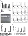
Figure 5
AmB-NA formulation-treated promastigote revealed reduced intracellular thiol and necrotic mode of cell death. (A) Effect of nanoparticles-based AmB-NA formulation as a function of the level of thiol derivative DTNB. Total intracellular reduced thiol was measured in deproteinized extracts of metacyclic L. donovani with or without drug formulation at the respective IC50 value of the drug formulation. After 12 h, AmB-NA (0.5 μM)-treated parasites revealed a ~3.2-fold decline in thiol release compared to AmB-treated parasites (p < 0.001). (B) Agarose (1%) gel electrophoresis of genomic DNA isolated from parasites treated with nanoparticles derived AmB-NA, AmB, AmBisome, and H2O2 with 100 bp and 1 kb DNA ladder as markers. Untreated and H2O2-treated (0.2 mM) parasites were taken as negative and positive controls, respectively. (C). A statistical representation of FACS data as a ratio of (%) of (PI+/AV+ cells) under similar conditions after 4 h and 8 h post-drug treatment. Promastigotes (1 × 10⁶ cells/mL) were treated with drugs at their IC50 and IC90 for 4 h and 8 h, respectively. Following 4 h incubation, the ratio was ~38 and ~8 for AmB-NA and AmB, respectively, whereas following 8 h incubation, it was ~42 and ~7, respectively. (D) Dot plot representation of FACS analysis corresponding to Annexin V and Propidium iodide-stained promastigotes after treatment with as-synthesized AmB-NA and commercially available AmB at their IC50 and IC90. (E) Evaluation of membrane leakage by LDH assay. A total of 1% Triton-X treated cells were measured as a positive control. Asterisks denote p values determined by one-way ANOVA analysis: *** p < 0.0001 compared with control, * p < 0.01 compared with the control. Analyzed data derived from at least 4 independent experiments * p < 0.05; ** 0.05 < p < 0.01; *** 0.01 < p < 0.001.
Figure 6
Figure 6
Effect of AmB-NA on the mitochondrial membrane potential of L. donovani promastigotes. The effect of AMB-NA formulation on L. donovani promastigotes mitochondrial membrane was determined. A known population of L. donovani promastigotes (1 × 10⁶/mL) was treated with as-synthesized AmB-NA for 4–8 h at 24 °C. The AmB-NA treated parasites were stained with JC-1 as described in the method. (A) FACS analysis of JC-1 mitochondrial potential marker staining 4 h and 8 h post-exposure with AmB-NA formulation. Panels represent density plots from a single analysis. Δψm depolarization monitored in the upper panel correspond to cells with intact mitochondrial membrane, whereas cells gated in the lower panel depicted damaged membrane of the treated cells with a loss of Δψm. (B) Graphic representation of mean values of the fluorescence corresponding to the mitochondrial membrane potential of the treated cells (Δψm collapse ± SD). (C) MFI was calculated employing a ratio of fluorescence at 590/530 nm. Asterisks (*) denote p values determined by one-way ANOVA analysis: *** p < 0.0001 compared with control, ** p < 0.001 compared with control, * p < 0.01 compared with the control. Analyzed data derived from at least 4 independent experiments.
Figure 7
Figure 7
Immunomodulatory potential of the AmB-NA in the L. donovani-infected BALB/c mice. The L. donovani-infected animals were treated with various as-synthesized AmB-NA formulations following a published protocol described in the method. The splenocytes of the cured animals were isolated after 4-week post-treatment as described in the method. (A) DTH response evoked in the immunized animal post-treatment with various forms of AmB. Footpad swelling was considered as a parameter to determine the as-generated cell-mediated immune response in the treated BALB/c mice. The treatments (50 μg SLA) were applied in a footpad, and the magnitude of DTH was assessed post 48 h. SLA was inoculated in the uninfected group, infected, untreated, and variously treated groups (AmB-NA (single dose), AmB-NA (double dose), and AmB). (B) Four-week post-treatment, blood from various experimental groups was collected to separate sera. ELISA was performed to evaluate antibody levels. IgG2a/IgG1 isotype ratios for AmB and AmB-NA single dose-treated mice were 1.9 and 2.1, respectively, thus proving AmB-NA to have better therapeutic efficacy against VL. Data represented means ± SE for mice per group and are representative of three independent experiments with similar results. ** p < 0.01, *** p < 0.001.
Figure 8
Figure 8
As-synthesized AmB-NA treatment resulted in successful clearance of the in vivo parasite burden. The treatment with AmB-NA resulted in the killing of the L. donovani systemic infection present in the experimental mice’s organs such as the spleen, liver, bone marrow, etc. Infected mice with no AmB treatment were considered as controls. The parasite burden in the spleen (A), liver (B), and bone marrow (C) was determined microscopically (Giemsa-stained cell smear) and expressed as no. of amastigotes/1000 cells. Data represented means ± SE for mice per group and are representative of three independent experiments with similar results. * p < 0.05, ** p < 0.01, *** p < 0.001, (D) The relative body weight of the experimental mice before and after drug administration. (E) Giemsa-stained uninfected (i), L. donovani-infected (ii) and L. donovani infected AmB-NA treated cells (iii). The arrows are showing the Ld bodies. Data represent the means ±SE for six animals per group. Results correspond to one of the three similar experiments.
Figure 9
Figure 9
Assessment of the pro-inflammatory potential of AmB-NA against murine visceral leishmaniasis. Four-week post-treatment splenocytes from different study groups (uninfected, infected, and various drug-treated BALB/c mice) were cultured and incubated in a CO2 incubator in the presence of 48 h SLA stimulation at 37 °C. Cell-free supernatant was used to evaluate cytokine profile through ELISA (A) IFN-γ, (B) IL-10, and (C). TNF-∝ (Data represented as means ± SE for six mice per group and are representative of two independent experiments with similar results. * p < 0.05, ** p < 0.01, *** p < 0.001), ns = non-significant, and (D) Semiquantitative PCR for genes specific for mice IFN-γ, IL-12, IL-10, and GAPDH.
Figure 10
Figure 10
Assessment of NO generation. The figure denotes nitric oxide (NO) released from macrophages belonging to various groups of untreated and treated mice (A) Quantitative estimation of NO produced (µM) in the cultured supernatant of the splenocytes. The freshly isolated mononuclear splenocytes cells were plated in a 12-well microtiter culture plate (1 × 10⁶ cells/mL/well) and incubated in the CO2 incubator at 37 °C for 48 h. Data represented means ± SE for six mice per group and are representative of three independent experiments with similar results. * p < 0.05, ** p < 0.01, *** p < 0.001, ns = non-significant. (B) Semi-quantitative PCR for genes specific for iNOS, GAPDH was used as a control. (C) Measurement of reactive oxygen species (ROS). Data represented means ± SE for six mice per group and are representative of three independent experiments with similar results. * p < 0.05, ** p < 0.01, *** p < 0.001.

Similar articles

Cited by

References

    1. Han S., Chen S.-B., Yang Z.-H., Feng Y., Wu W.-P. Epidemiology of Leishmania Carriers in Tan Chang County, Gansu Province, China. Front. Cell. Infect. Microbiol. 2021;11:645944. doi: 10.3389/fcimb.2021.645944. - DOI - PMC - PubMed
    1. Torres-Guerrero E., Quintanilla-Cedillo M.R., Ruiz-Esmenjaud J., Arenas R. Leishmaniasis: A review. F1000Research. 2017;6:750. doi: 10.12688/f1000research.11120.1. - DOI - PMC - PubMed
    1. Rijal S., Sundar S., Mondal D., Das P., Alvar J., Boelaert M. Eliminating visceral leishmaniasis in South Asia: The road ahead. BMJ. 2019;364:k5224. doi: 10.1136/bmj.k5224. - DOI - PMC - PubMed
    1. Akuffo H., Costa C., Van Griensven J., Burza S., Moreno J., Herrero M. New insights into leishmaniasis in the immunosuppressed. PLOS Negl. Trop. Dis. 2018;12:e0006375. doi: 10.1371/journal.pntd.0006375. - DOI - PMC - PubMed
    1. Leishmaniasis, WHO 2021. [(accessed on 19 October 2021)]. Available online: https://www.who.int/news-room/fact-sheets/detail/leishmaniasis.

LinkOut - more resources