Review
. 2022 Dec 21;15(1):32.
doi: 10.3390/v15010032.

Determinants of Retroviral Integration and Implications for Gene Therapeutic MLV-Based Vectors and for a Cure for HIV-1 Infection

Affiliations
Review

Determinants of Retroviral Integration and Implications for Gene Therapeutic MLV-Based Vectors and for a Cure for HIV-1 Infection

Eline Pellaers et al. Viruses. .

Abstract

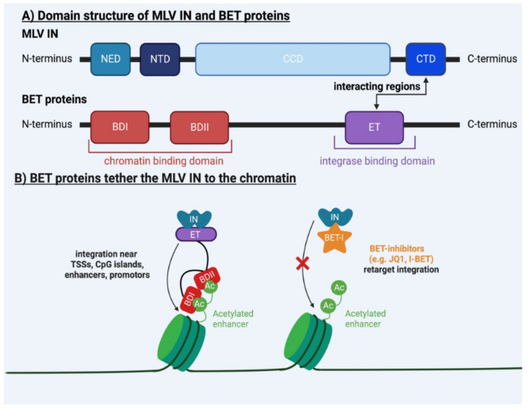
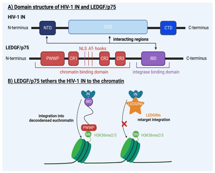
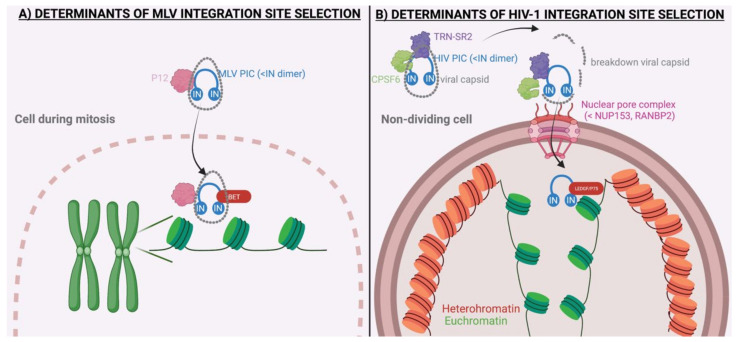
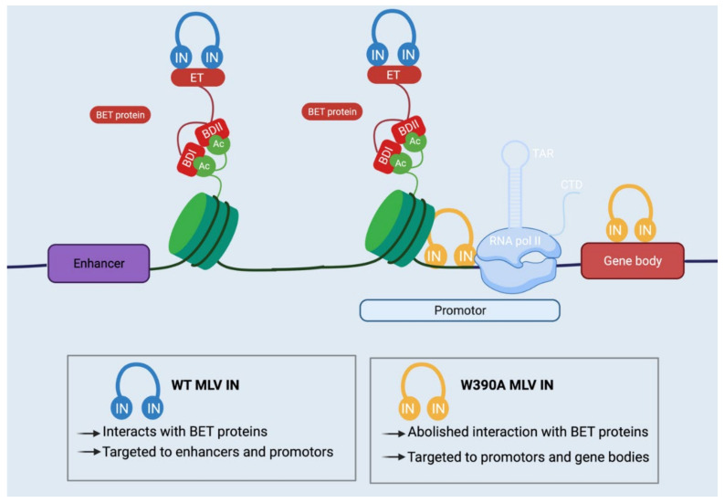
To complete their replication cycle, retroviruses need to integrate a DNA copy of their RNA genome into a host chromosome. Integration site selection is not random and is driven by multiple viral and cellular host factors specific to different classes of retroviruses. Today, overwhelming evidence from cell culture, animal experiments and clinical data suggests that integration sites are important for retroviral replication, oncogenesis and/or latency. In this review, we will summarize the increasing knowledge of the mechanisms underlying the integration site selection of the gammaretrovirus MLV and the lentivirus HIV-1. We will discuss how host factors of the integration site selection of retroviruses may steer the development of safer viral vectors for gene therapy. Next, we will discuss how altering the integration site preference of HIV-1 using small molecules could lead to a cure for HIV-1 infection.

Keywords: HIV-1; MLV; host factors; integration site selection; latency; retroviral integration; viral vector.


Conflict of interest statement

The authors declare no conflict of interest.

Figures

Figure 1
Figure 1
(A) Domain structure of murine leukemia virus (MLV) integrase (IN) and bromodomain and extra–terminal motif (BET) proteins. The MLV IN consists of an N–terminal extension domain (NED), an N–terminal domain (NTD), a catalytic core domain (CCD) and a C–terminal domain (CTD) [4]. BET proteins consist of two bromodomains (BD), BDI and BDII, that bind acetylated lysines in the nucleosomes, and an extra–terminal (ET) domain that interacts with the CTD of the MLV IN [28,29,30,31,32,33,34]. (B) BET proteins tether the MLV IN. Through the combined interaction of the ET domain of BET proteins (which binds the CTD of the MLV IN) and BDI and BDII (which bind the acetylated chromatin), BET proteins tether the MLV IN to acetylated chromatin regions such as enhancers [28,29,30,31,32,33,34]. BET inhibitors, such as JQ1 and I–BET, retarget the integration of MLV away from BET–recognized sites by uncoupling the interaction between BET proteins and MLV IN [29,30,31]. (Figure created with Biorender.com on 17 December 2022).
Figure 2
Figure 2
(A) Domain structure of HIV-1 integrase (IN) and lens epithelium–derived growth factor (LEDGF/p75). HIV-1 IN consists of an N–terminal domain (NTD), a catalytic core domain (CCD) and a C–terminal domain (CTD) [3]. LEDGF/p75 consists of an integrase–binding domain (IBD), binding the viral IN, and a PWWP domain, binding epigenetic marks in the genome associated with active transcription such as H3K36me2/3 [38,39,40,41,42]. (B) LEDGF/p75 tethers HIV-1 IN to chromatin. LEDGF/p75 simultaneously binds HIV-1 IN with its integrase–binding domain (IBD) and chromatin through its PWWP domain, tethering HIV-1 IN to decondensed chromatin regions, tagged by H3K36me2/3 [38,39,40,41,42]. HIV-1 integration can be targeted away from H3K36me2/3 with the use of LEDGINs, small molecules that inhibit the interaction between HIV-1 IN and LEDGF/p75 [22,23]. (Figure created with Biorender.com on 17 December 2022).
Figure 3
Figure 3
(A) Determinants of the murine leukemia virus (MLV). MLV tends to integrate near transcription start sites (TSSs) and enhancers. The MLV p12 functions as a tether of the pre–integration complex (PIC) to the chromosomes, but its role in integration site selection is under debate. In contrast to HIV-1, MLV does not have an active nuclear entry route but is dependent on mitosis for nuclear entry. After nuclear entry and mitosis, bromodomain and extra–terminal motif (BET) proteins direct the integration pattern of MLV. BET proteins consist of an extra–terminal domain, which binds the MLV integrase (IN), and two bromodomains (BDI and BDII) that bin acetylated lysine in the nucleosomes. (B) Determinants of human immunodeficiency virus 1 (HIV-1). HIV-1 tends to integrate near decondensed chromatin, associated with active transcription, at the nuclear periphery. The HIV-1 pre–integration complex (PIC) can infect non–dividing cells by crossing the intact nuclear membrane via nuclear pore complexes. Nuclear import affects the HIV-1 integration pattern, with the ran–binding protein 2 (RANBP2, also called Nup358), transportin SR2 (TRN–SR2, also called transportin 3) and nucleoporin 153 (Nup153) as determinants of integration site selection. After nuclear import, lens epithelium–derived growth factor (LEDGF/p75) is considered the major contributing factor to integration site selection. LEDGF/p75 consists of an integrase–binding domain (IBD), binding the viral IN, and a PWWP domain, binding epigenetic marks in the genome associated with active transcription such as H3K36me2/3. In addition, the three–dimensional organization in the nucleus and the epigenetic landscape of chromatin affect HIV integration, as HIV-1 tends to integrate into less condensed euchromatin and avoids integration in closed heterochromatin regions. (Figure created with Biorender.com on 17 December 2022).
Figure 4
Figure 4
Integration pattern of wild–type (WT) and W380A mutant murine leukemia virus (MLV) in vivo. Bromodomain and extra–terminal motif (BET) proteins tether the wild–type (WT) MLV pre–integration complex (PIC) to enhancers and promoters. In contrast, integration of the W390A mutant MLV PIC, which cannot interact with BET, is redirected toward promoters and gene bodies. Oncogenesis by WT MLV IN is probably provoked by integration near enhancers and promotors, which results in the overexpression of cellular oncogenes, while the W390A mutant’s integration in oncogene bodies induces insertional mutagenesis [55] (Figure created with Biorender.com on 17 December 2022).

Similar articles

Cited by

References

    1. Greenwood A.D., Ishida Y., O’Brien S.P., Roca A.L., Eiden M.V. Transmission, Evolution, and Endogenization: Lessons Learned from Recent Retroviral Invasions. Microbiol. Mol. Biol. Rev. 2018;82:e00044-17. doi: 10.1128/MMBR.00044-17. - DOI - PMC - PubMed
    1. Nisole S., Saïb A. Early Steps of Retrovirus Replicative Cycle. Retrovirology. 2004;1:1–20. doi: 10.1186/1742-4690-1-9. - DOI - PMC - PubMed
    1. Passos D.O., Li M., Craigie R., Lyumkis D. Retroviral Integrase: Structure, Mechanism, and Inhibition. Enzymes. 2022;50:249–300. doi: 10.1016/bs.enz.2021.06.007. - DOI - PMC - PubMed
    1. Cherepanov P., Maertens G.N., Hare S. Structural Insights into the Retroviral DNA Integration Apparatus. Curr. Opin. Struct. Biol. 2011;21:249–256. doi: 10.1016/j.sbi.2010.12.005. - DOI - PubMed
    1. Barr S.D., Leipzig J., Shinn P., Ecker J.R., Bushman F.D. Integration Targeting by Avian Sarcoma–Leukosis Virus and Human Immunodeficiency Virus in the Chicken Genome. J. Virol. 2005;79:12035–12044. doi: 10.1128/JVI.79.18.12035-12044.2005. - DOI - PMC - PubMed

Publication types